Phẫu thuật tiến hai hàm trong điều trị ngưng thở khi ngủ tắc nghẽnMaxillomandibular advancement for obstruCTive sleep apnea
02/09/2026Kết quảResults
Ngưng thở khi ngủ tắc nghẽn là một rối loạn hô hấp trong khi ngủ, trong đó đường thở trên bị hẹp hoặc xẹp lặp đi lặp lại, gây ra các đợt giảm hoặc ngừng luồng khí dù cơ thể vẫn tiếp tục nỗ lực hô hấp.
Biểu hiện thường gặp bao gồm ngủ ngáy, những khoảng ngừng thở được người bên cạnh quan sát thấy, giấc ngủ không phục hồi, buồn ngủ ban ngày, mệt mỏi và giảm khả năng tập trung.
Ở một số bệnh nhân, cấu trúc xương hàm và khuôn mặt đóng vai trò quan trọng trong sự hẹp của đường thở. Khi đó, phẫu thuật chỉnh hình xương hàm không chỉ nhằm thay đổi khớp cắn hoặc thẩm mỹ khuôn mặt mà còn có thể trở thành một phương pháp điều trị ngưng thở khi ngủ.
Khi cấu trúc khuôn mặt liên quan đến đường thở
Bệnh nhân trong trường hợp này đến khám sau một thời gian dài ngủ ngáy lớn, thường xuyên buồn ngủ, mệt mỏi và khó tập trung vào ban ngày.
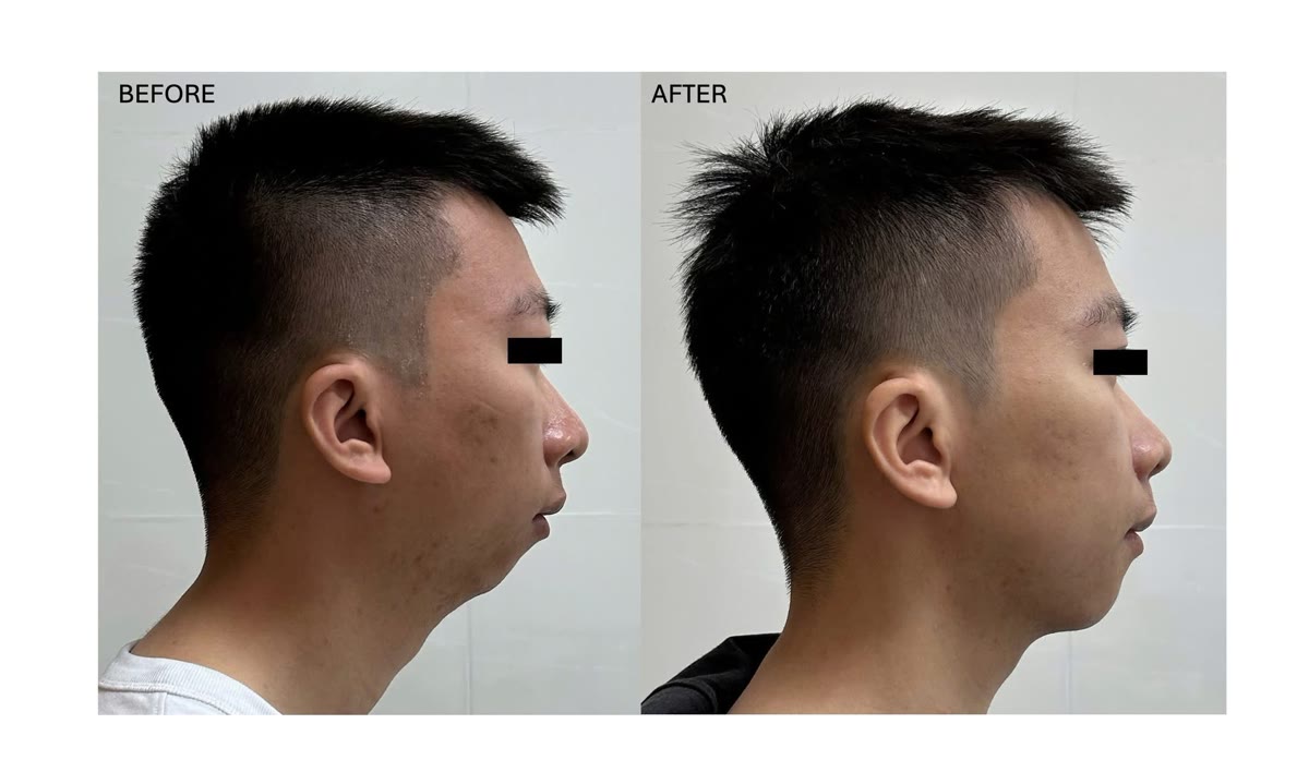
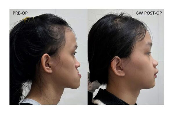
Khám lâm sàng ghi nhận hàm dưới nhỏ và lùi, cằm kém nhô và góc cằm – cổ tù. Bệnh nhân cũng có xu hướng giữ đầu hơi đưa ra trước.
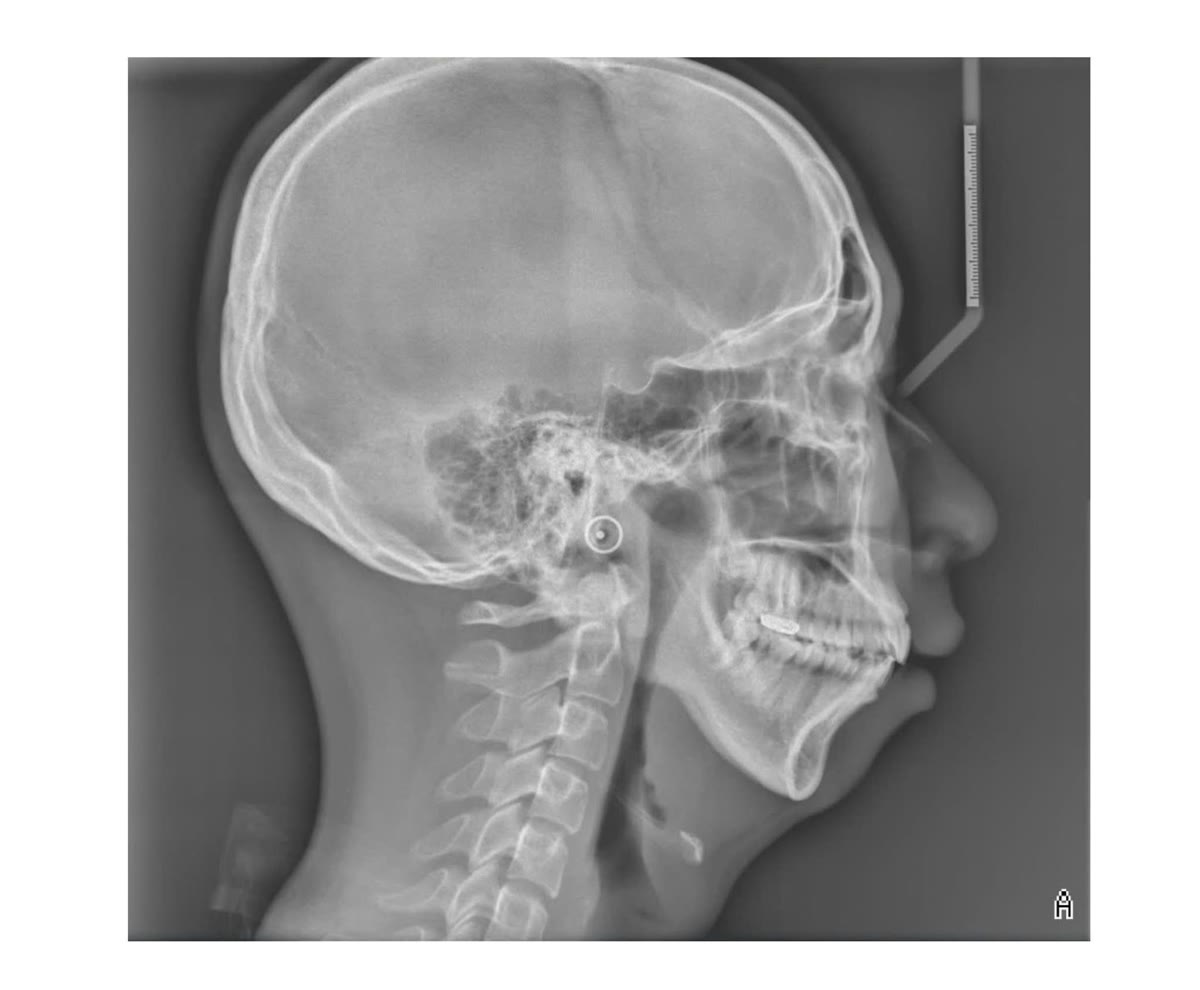
Trên hình ảnh sọ mặt, khoảng đường thở phía sau tương đối hẹp.
Tuy nhiên, hình ảnh đường thở đơn thuần không thể chẩn đoán ngưng thở khi ngủ.
Bệnh nhân được thực hiện đa ký giấc ngủ và ghi nhận chỉ số ngưng thở – giảm thở AHI là 47 lần mỗi giờ, tương ứng với ngưng thở khi ngủ tắc nghẽn mức độ nặng.
Việc kết hợp đánh giá giấc ngủ với phân tích cấu trúc sọ mặt cho phép xây dựng một kế hoạch điều trị phù hợp hơn cho từng bệnh nhân.
Tại sao tiến hai hàm có thể cải thiện đường thở?
Phẫu thuật tiến hai hàm là một trong những phương pháp phẫu thuật được sử dụng để điều trị ngưng thở khi ngủ tắc nghẽn.
Khác với những phẫu thuật chỉ tác động vào một vị trí của đường thở, phương pháp này thay đổi cấu trúc nâng đỡ của toàn bộ vùng hầu bằng cách đưa xương hàm trên và hàm dưới ra trước.
Khi hai hàm được tiến, các mô mềm liên quan đến khẩu cái, lưỡi và vùng trên móng cũng được đưa ra trước theo hệ thống điểm bám của chúng. Nhờ đó, không gian đường thở phía sau có thể được mở rộng và khả năng xẹp của đường thở trong khi ngủ có thể giảm.
Đây là lý do phương pháp này đặc biệt đáng cân nhắc ở những bệnh nhân có OSA đi kèm cấu trúc xương hàm lùi.
Không chỉ là “đưa hai hàm ra trước”
Ở bệnh nhân này, kế hoạch phẫu thuật không đơn thuần là tịnh tiến hai hàm theo chiều trước – sau.
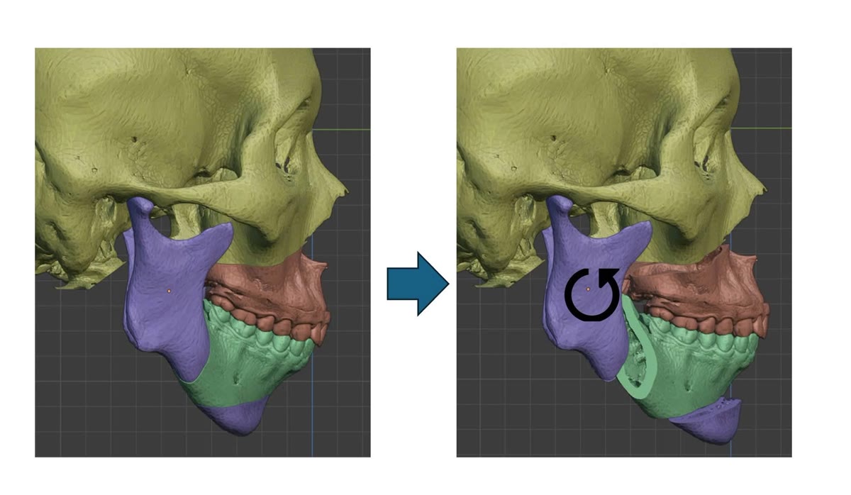
Xương hàm trên và hàm dưới được đưa ra trước kết hợp xoay phức hợp hai hàm ngược chiều kim đồng hồ.
Kiểu di chuyển này cho phép tăng mức độ đưa ra trước của hàm dưới và vùng cằm, đồng thời kiểm soát tương quan khớp cắn, chiều cao tầng mặt và hình thể khuôn mặt.
Tạo hình cằm được thực hiện đồng thời để hoàn thiện tương quan cằm – môi – cổ và sự cân đối của tầng mặt dưới.
Điều quan trọng là lượng tiến và mức độ xoay không thể áp dụng theo một công thức cố định.
Mỗi bệnh nhân cần được đánh giá đồng thời về đường thở – khớp cắn – cấu trúc xương – mô mềm khuôn mặt trước khi quyết định vị trí cuối cùng của hai hàm.
Lập kế hoạch phẫu thuật ba chiều
Toàn bộ phẫu thuật được mô phỏng trước trên mô hình ba chiều của bệnh nhân.
Vị trí ban đầu của xương hàm, tương quan khớp cắn và hình thái khuôn mặt được phân tích trước khi xác định hướng và mức độ di chuyển của từng đoạn xương.
Các dụng cụ hướng dẫn phẫu thuật cá nhân hóa sau đó được sử dụng để chuyển kế hoạch từ môi trường số vào phòng mổ.
Đối với phẫu thuật điều trị OSA, lập kế hoạch ba chiều đặc biệt hữu ích vì mục tiêu không chỉ là đạt được một khớp cắn phù hợp.
Kế hoạch cần đồng thời tạo ra một thay đổi có lợi cho đường thở nhưng vẫn duy trì sự hài hòa của khuôn mặt.
Kết quả sau sáu tháng
Ở thời điểm sáu tháng sau phẫu thuật, bệnh nhân ghi nhận sự thay đổi rõ rệt về chất lượng giấc ngủ.
Tình trạng ngủ ngáy giảm rất nhiều, giấc ngủ sâu và phục hồi tốt hơn. Ban ngày bệnh nhân tỉnh táo hơn, giảm mệt mỏi và cải thiện khả năng tập trung trong học tập và công việc.
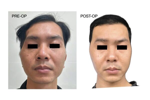
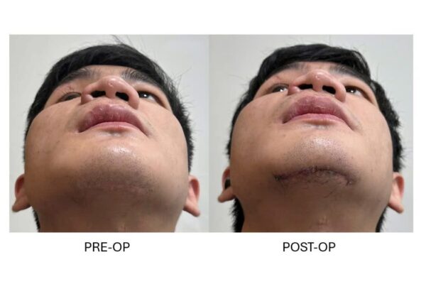
Đồng thời, việc đưa hàm dưới và cằm ra trước giúp cải thiện rõ nét hình thể tầng mặt dưới, tương quan môi – cằm và đường viền cằm – cổ.
Những thay đổi chủ quan này rất có ý nghĩa đối với người bệnh. Tuy nhiên, trong điều trị OSA, cải thiện triệu chứng không thể thay thế cho đánh giá khách quan bằng nghiên cứu giấc ngủ sau phẫu thuật. Theo dõi lâu dài vẫn cần thiết để đánh giá đầy đủ mức độ kiểm soát bệnh.
MMA là một trong những phẫu thuật hiệu quả nhất đối với OSA được lựa chọn phù hợp
Phẫu thuật tiến hai hàm có thể tạo ra mức giảm AHI đáng kể ở nhiều bệnh nhân và được xem là một trong những phương pháp phẫu thuật có hiệu quả cao trong điều trị OSA.
Tuy nhiên, “thành công” và “khỏi bệnh” là hai khái niệm khác nhau.
Trong các nghiên cứu về OSA, thành công phẫu thuật thường được định nghĩa dựa trên mức giảm AHI và một ngưỡng AHI sau điều trị, trong khi khỏi bệnh đòi hỏi tiêu chuẩn nghiêm ngặt hơn. Vì vậy, không nên diễn giải một tỷ lệ thành công thành khả năng chữa khỏi hoàn toàn cho mọi bệnh nhân.
Kết quả cũng phụ thuộc vào nhiều yếu tố như mức độ bệnh, cấu trúc sọ mặt, cân nặng, tuổi, mức độ tiến xương, vị trí tắc nghẽn và các yếu tố giải phẫu khác.
Phẫu thuật chỉnh hình xương hàm không chỉ điều trị khớp cắn
Trường hợp này cho thấy một khía cạnh khác của phẫu thuật chỉnh hình xương hàm.
Ở những bệnh nhân phù hợp, vị trí của xương hàm không chỉ ảnh hưởng đến khớp cắn và hình thể khuôn mặt mà còn liên quan đến cấu trúc nâng đỡ đường thở.
Vì vậy, một kế hoạch phẫu thuật tốt cần nhìn bệnh nhân dưới nhiều góc độ:
khớp cắn – khuôn mặt – đường thở – giấc ngủ.
Mục tiêu cuối cùng không chỉ là tạo ra một khuôn mặt cân đối hơn, mà còn là cải thiện chức năng và chất lượng sống của người bệnh.
Obstructive sleep apnea (OSA) is a sleep-related breathing disorder characterized by recurrent narrowing or collapse of the upper airway, resulting in repeated episodes of reduced or absent airflow despite ongoing respiratory effort.
Common manifestations include loud snoring, witnessed apneas, non-restorative sleep, excessive daytime sleepiness, fatigue, and impaired concentration.
In selected patients, craniofacial anatomy contributes substantially to upper-airway restriction. In this setting, orthognathic surgery can serve not only to modify occlusion and facial appearance, but also as a surgical treatment for OSA.
When craniofacial anatomy contributes to airway obstruction
The patient in this case presented with a history of severe snoring, daytime sleepiness, fatigue, and difficulty concentrating.
Clinical examination demonstrated mandibular deficiency and retrusion, limited chin projection, and an obtuse cervicomental relationship. The patient also tended to maintain a slightly forward head posture.
Craniofacial imaging demonstrated a relatively narrow posterior airway space.
Imaging alone, however, cannot establish the diagnosis or severity of OSA.
Polysomnography demonstrated an apnea–hypopnea index (AHI) of 47 events per hour, consistent with severe obstructive sleep apnea.
Combining objective sleep assessment with craniofacial analysis allowed the treatment strategy to address both the sleep disorder and the patient's underlying skeletal anatomy.
Why can maxillomandibular advancement improve the airway?
Maxillomandibular advancement (MMA) is an established surgical option for the treatment of obstructive sleep apnea.
Unlike procedures targeting a single anatomical level of obstruction, MMA modifies the skeletal framework supporting multiple levels of the upper airway by advancing both the maxilla and mandible.
Advancement of the jaws carries forward associated soft-tissue attachments involving the palate, tongue, and suprahyoid complex. This can increase upper-airway dimensions and reduce its tendency to collapse during sleep.
For this reason, MMA is particularly relevant in appropriately selected patients with OSA and retrusive craniofacial anatomy.
More than simply moving both jaws forward
In this patient, the surgical plan involved more than straightforward anteroposterior advancement.
The maxillomandibular complex was advanced and underwent counterclockwise rotation.
This movement pattern allowed substantial advancement of the mandible and chin while simultaneously controlling the occlusal relationship, vertical facial proportions, and overall facial profile.
Genioplasty was performed concurrently to further optimize chin projection, lower facial balance, and the cervicomental contour.
Importantly, there is no universal amount of advancement or rotation that can be applied to every patient.
Planning requires simultaneous assessment of the airway, occlusion, skeletal framework, and facial soft tissues before determining the final position of the jaws.
Three-dimensional surgical planning
The operation was planned preoperatively using a three-dimensional model of the patient's craniofacial anatomy.
The initial skeletal position, occlusal relationship, and facial morphology were analyzed before determining the direction and magnitude of movement for each skeletal segment.
Patient-specific surgical guidance was then used to transfer the virtual plan to the operating room.
In OSA surgery, three-dimensional planning is particularly valuable because obtaining an appropriate occlusion is only one component of the treatment objective.
The surgical plan must also create a favorable airway change while maintaining or improving facial harmony.
Six-month outcome
At six months after surgery, the patient reported a substantial improvement in sleep quality.
Snoring had markedly decreased, sleep was deeper and more restorative, and daytime alertness and concentration had improved.
Advancement of the mandibular and chin framework also produced a significant change in lower facial projection, lip–chin balance, and cervicomental contour.
These subjective improvements are clinically meaningful. However, in OSA treatment, symptomatic improvement does not replace objective postoperative sleep assessment. Long-term follow-up and repeat sleep testing remain important for determining the degree of disease control.
MMA is one of the most effective surgical treatments for appropriately selected patients with OSA
Maxillomandibular advancement can produce substantial reductions in AHI and is regarded as one of the most effective surgical options for appropriately selected patients with obstructive sleep apnea.
However, surgical “success” and “cure” should not be considered synonymous.
Studies of OSA surgery commonly define surgical success according to a specified reduction in AHI combined with a postoperative AHI threshold, whereas cure or remission requires more stringent criteria. A reported surgical success rate should therefore not be interpreted as a universal probability of complete elimination of OSA.
Outcomes also depend on multiple factors, including baseline disease severity, craniofacial anatomy, body weight, age, magnitude of skeletal advancement, sites of airway obstruction, and other anatomical and physiological factors.
Orthognathic surgery is not only about occlusion
This case illustrates a broader role of orthognathic surgery.
In appropriately selected patients, jaw position influences not only dental occlusion and facial appearance but also the skeletal support of the upper airway.
A comprehensive surgical plan should therefore consider four interconnected objectives:
occlusion – facial aesthetics – airway – sleep.
The ultimate goal is not simply to create a more balanced facial profile, but to improve function, sleep, and quality of life.