Tái tạo lõm thái dương sau tiêu xương sọ bằng mảnh ghép peek cá nhân hóaPatient-specific PEEK cranioplasty for postoperative temporal hollowing
02/09/2026Kết quảResults
Tái tạo khuyết hổng hộp sọ không chỉ là phục hồi phần xương bị mất. Ở những vùng có ảnh hưởng trực tiếp đến đường nét khuôn mặt, đặc biệt là vùng thái dương, hình dạng của mảnh ghép cần được thiết kế dựa trên cả xương và thể tích mô mềm bên ngoài.
Trường hợp này là một ví dụ về việc ứng dụng thiết kế ba chiều và mảnh ghép PEEK cá nhân hóa để giải quyết đồng thời hai vấn đề: khuyết hổng xương sọ và lõm thái dương sau phẫu thuật.
Khi mảnh xương sọ được ghép lại nhưng sau đó bị tiêu
Bệnh nhân từng bị chấn thương sọ não và phải phẫu thuật mở sọ giải áp. Trong phẫu thuật này, một phần xương sọ được tạm thời lấy bỏ nhằm tạo thêm không gian cho não trong giai đoạn phù nề.
Sau đó, mảnh xương sọ được ghép trở lại.
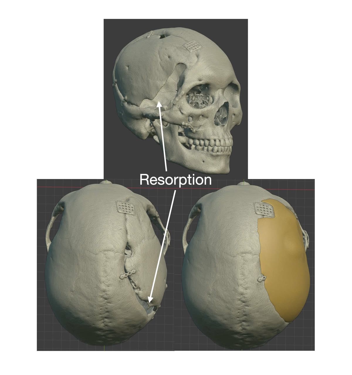
Tuy nhiên, theo thời gian, một phần xương vùng thái dương bên phải bị tiêu, tạo ra khuyết hổng và làm thay đổi cấu trúc nâng đỡ của vùng thái dương.
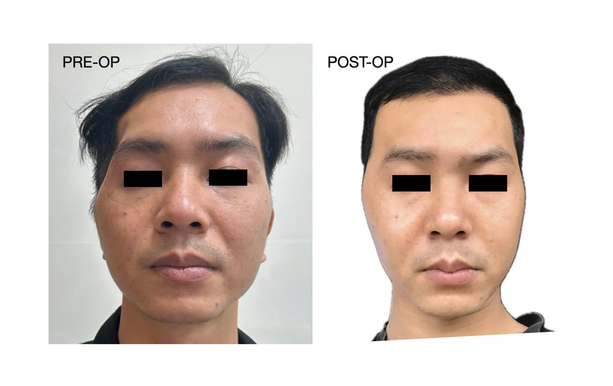
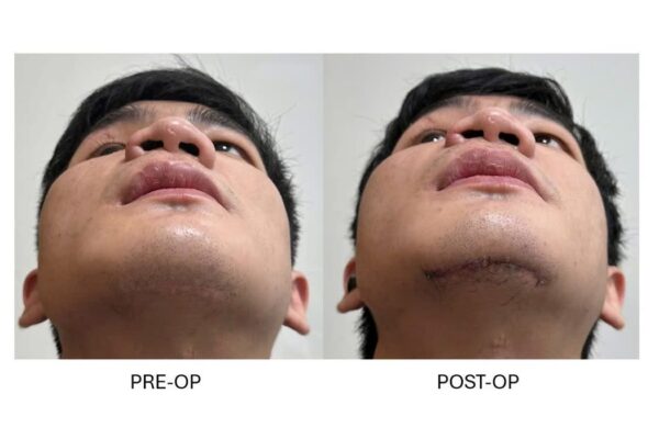
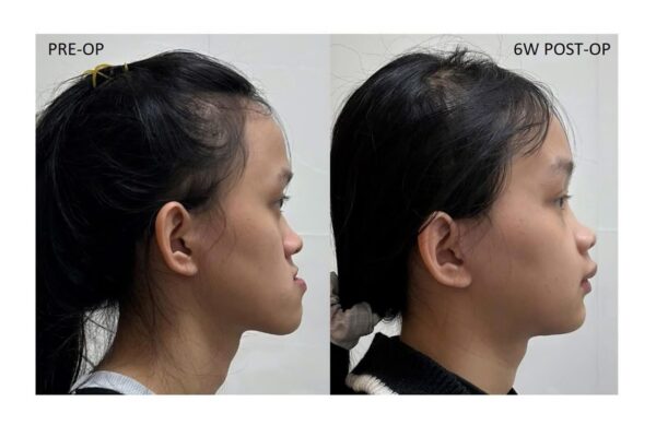
Hậu quả dễ nhận thấy nhất là lõm thái dương, tạo ra sự khác biệt rõ rệt giữa hai bên khuôn mặt.
Lõm thái dương không đơn thuần là một khuyết hổng xương
Hình thể vùng thái dương được tạo nên bởi sự kết hợp giữa xương sọ, cơ thái dương, lớp mỡ và các mô mềm bao phủ.
Sau chấn thương hoặc phẫu thuật sọ não, cơ thái dương có thể bị teo, mất hoặc thay đổi điểm bám, thay đổi vị trí và tương quan với nền xương bên dưới. Nếu đồng thời xảy ra tiêu xương sọ, cấu trúc nâng đỡ cơ và mô mềm càng bị thay đổi.
Ở bệnh nhân này, mặc dù cơ thái dương đã được xử lý cẩn thận trong lần phẫu thuật trước, sự tiêu xương vùng thái dương làm mất một phần nền nâng đỡ, khiến cơ và mô mềm dịch vào phía trong và tạo nên vùng lõm rõ rệt.
Điều này đặt ra một vấn đề quan trọng:
Nếu chỉ tái tạo chính xác phần xương bị mất, liệu đường nét bên ngoài của vùng thái dương có thực sự được phục hồi?
Không phải lúc nào câu trả lời cũng là có.
Thiết kế mảnh ghép dựa trên cả xương và mô mềm
Một số phương pháp tăng thể tích mô mềm như ghép mỡ tự thân có thể được sử dụng để điều trị lõm thái dương. Tuy nhiên, mức độ duy trì thể tích của mỡ ghép có thể thay đổi và một số bệnh nhân cần thực hiện bổ sung.
Trong trường hợp này, một chiến lược khác được lựa chọn: sử dụng chính hình thể bên lành của bệnh nhân để xác định thể tích cần tái tạo.
Đầu tiên, dữ liệu hình ảnh của bệnh nhân được sử dụng để dựng lại mô hình ba chiều của hộp sọ. Mô hình này được gửi đến đơn vị chế tạo để xây dựng thiết kế ban đầu của mảnh ghép hộp sọ cá nhân hóa.
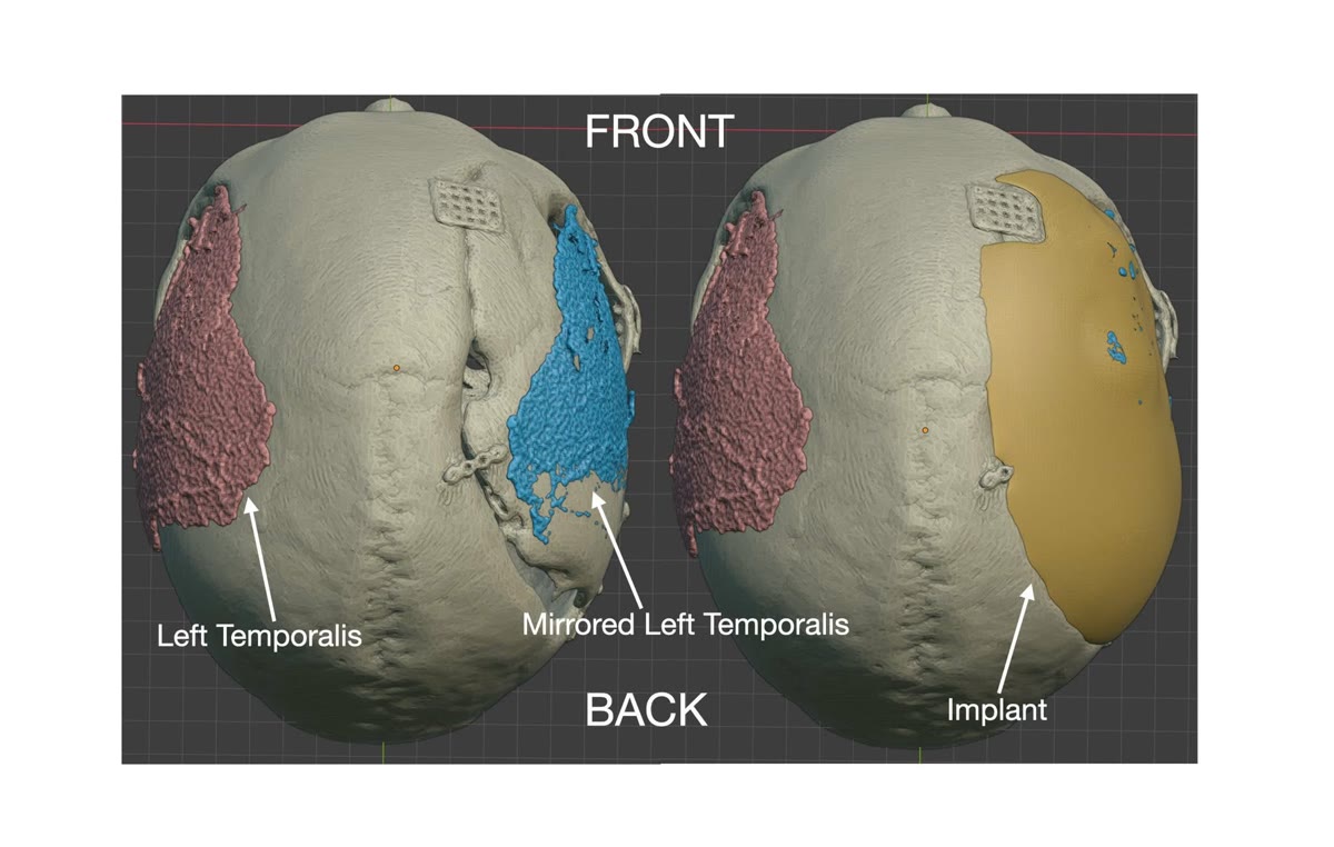
Song song với đó, thể tích của cơ thái dương bên lành được dựng lại trên phần mềm Blender.
Cấu trúc này sau đó được đối xứng sang bên tổn thương, tạo ra một tham chiếu ba chiều về hình thể vùng thái dương mà chúng ta muốn tái lập.
Mảnh ghép không chỉ thay thế phần xương bị mất
Nếu mảnh ghép chỉ được thiết kế theo đúng bề mặt xương sọ ban đầu, phần thiếu hụt về cơ và mô mềm phía ngoài có thể khiến vùng thái dương vẫn bị lõm sau phẫu thuật.
Vì vậy, thiết kế ban đầu được chỉnh sửa.
Ở những vùng cần thiết, mảnh ghép được làm dày có chủ đích dựa trên thể tích mô mềm của bên lành đã được đối xứng sang.
Nói cách khác, mục tiêu thiết kế không còn đơn thuần là:
“Phần xương nào đang bị thiếu?”
mà trở thành:
“Cần tái tạo bao nhiêu thể tích để đường nét bên ngoài của hai bên trở nên cân đối hơn?”
Sau khi hoàn thiện, thiết kế được gửi lại cho đơn vị chế tạo và mảnh ghép cá nhân hóa được sản xuất bằng PEEK (polyetheretherketone).
Phối hợp phẫu thuật thần kinh và hàm mặt
Ca phẫu thuật được thực hiện với sự phối hợp giữa phẫu thuật thần kinh và phẫu thuật hàm mặt.
Sau khi bộc lộ vùng khuyết hổng, cơ thái dương và các mô xung quanh được bóc tách và sắp xếp lại để phù hợp với hình thể mới.
Mảnh ghép PEEK cá nhân hóa sau đó được đặt vào vị trí để đồng thời tái tạo khuyết hổng hộp sọ và tạo lại nền nâng đỡ cho vùng thái dương.
Trong cùng cuộc phẫu thuật, biến dạng còn lại của vùng xương gò má cũng được điều chỉnh nhằm cải thiện sự liên tục của đường nét vùng thái dương – gò má.
Kết quả sau ba tháng
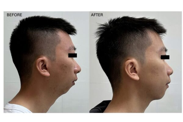
Sau ba tháng, sự cân đối của vùng thái dương được cải thiện rõ rệt.
Thể tích mô mềm bên tổn thương được phục hồi tốt hơn và đường chuyển tiếp từ vùng thái dương xuống gò má trở nên tự nhiên hơn.
Điểm quan trọng của kết quả này không chỉ nằm ở việc khuyết hổng xương sọ được che phủ, mà ở việc hình dạng bên ngoài của khuôn mặt đã được đưa vào thiết kế ngay từ trước phẫu thuật.
Xương tự thân hay vật liệu nhân tạo?
Trong tái tạo sọ mặt, không có một vật liệu duy nhất phù hợp cho tất cả các khuyết hổng.
Xương tự thân có nhiều ưu điểm về mặt sinh học và có khả năng tích hợp với hệ xương của bệnh nhân. Tuy nhiên, xương ghép cũng có thể bị tiêu theo thời gian, đặc biệt trong một số tình huống tái tạo hộp sọ.
Các vật liệu nhân tạo như PEEK không có khả năng tạo xương như mô tự thân, nhưng có ưu điểm là duy trì hình dạng đã được thiết kế và có thể chế tạo chính xác theo hình thái giải phẫu riêng của từng bệnh nhân.
Do đó, lựa chọn giữa xương tự thân và vật liệu nhân tạo cần dựa trên vị trí, kích thước và đặc điểm của khuyết hổng, tình trạng mô mềm, tiền sử phẫu thuật cũng như mục tiêu chức năng và thẩm mỹ của từng trường hợp.
Từ tái tạo khuyết hổng đến tái tạo hình thể
Trường hợp này minh họa một thay đổi quan trọng trong cách tiếp cận tái tạo sọ mặt.
Với công nghệ thiết kế ba chiều, câu hỏi không còn chỉ là làm thế nào để tạo ra một mảnh ghép vừa với phần xương bị thiếu.
Chúng ta có thể sử dụng dữ liệu giải phẫu của chính bệnh nhân để thiết kế hình thể cuối cùng mà mình muốn tái tạo.
Ở vùng thái dương, điều đó đặc biệt quan trọng bởi đường nét bên ngoài không chỉ được quyết định bởi xương mà còn bởi cơ và mô mềm.
Vì vậy, một mảnh ghép cá nhân hóa đôi khi cần được thiết kế không giống hoàn toàn phần xương đã mất, mà được điều chỉnh có chủ đích để bù lại những thay đổi của mô mềm bên ngoài.
Đó cũng là một trong những giá trị lớn nhất của thiết kế ba chiều trong phẫu thuật tái tạo: không chỉ tái tạo cấu trúc đã mất, mà còn cho phép thiết kế kết quả cần đạt được cho từng bệnh nhân.
Cranial reconstruction is not simply about replacing missing bone. In regions that directly influence facial contour, particularly the temporal area, the implant may need to be designed according to both the skeletal defect and the overlying soft-tissue volume.
This case demonstrates the use of three-dimensional planning and a patient-specific PEEK implant to address two related problems simultaneously: a cranial defect and postoperative temporal hollowing.
When the replaced cranial bone subsequently resorbs
The patient had previously sustained a traumatic brain injury requiring decompressive craniectomy. During this procedure, a portion of the skull was temporarily removed to accommodate cerebral swelling.
The cranial bone flap was subsequently replaced.
Over time, however, part of the right temporal bone underwent resorption, resulting in a secondary cranial defect and loss of structural support in the temporal region.
The most visible consequence was significant temporal hollowing, producing a clear asymmetry between the two sides of the face.
Temporal hollowing is not simply a bony defect
The contour of the temporal region is determined by the combined anatomy of the cranial skeleton, temporalis muscle, fat, and overlying soft tissues.
Following trauma or cranial surgery, the temporalis muscle may undergo atrophy, loss or alteration of its attachment, displacement, or changes in its relationship with the underlying skeleton. When cranial bone resorption occurs simultaneously, the structural support for the muscle and soft tissues may be further compromised.
In this patient, despite careful management of the temporalis during the previous procedure, resorption of the temporal bone removed part of its underlying support, allowing the muscle and surrounding tissues to displace medially and creating a pronounced temporal depression.
This raises an important reconstructive question:
If we simply replace the missing bone anatomically, will the external temporal contour also be restored?
Not necessarily.
Designing the reconstruction around both bone and soft tissue
Soft-tissue augmentation techniques such as autologous fat grafting can be used for temporal hollowing. However, retention of grafted fat volume can vary, and additional procedures may occasionally be required.
For this patient, a different strategy was selected: the unaffected side of the patient's own anatomy was used as the reference for the volume to be reconstructed.
First, the patient's imaging data were used to reconstruct a three-dimensional model of the skull. This model was provided to the manufacturer to generate the initial design of a patient-specific cranial implant.
In parallel, the volume of the unaffected temporalis muscle was reconstructed using Blender.
This volume was then mirrored to the affected side, providing a patient-specific three-dimensional reference for the desired temporal contour.
An implant designed to do more than replace missing bone
If the implant had simply reproduced the original bony surface, the loss of muscle and soft-tissue volume could have resulted in persistent temporal depression despite successful reconstruction of the cranial defect.
The initial implant design was therefore modified.
In selected areas, the implant was intentionally augmented beyond the reconstructed skeletal contour, using the mirrored soft-tissue volume from the unaffected side as a reference.
The design question therefore changed from:
“What bone is missing?”
to:
“What volume needs to be reconstructed to restore symmetry of the external contour?”
Once the design was finalized, the patient-specific implant was manufactured from PEEK (polyetheretherketone).
Collaboration between neurosurgery and maxillofacial surgery
The operation was performed through collaboration between neurosurgery and maxillofacial surgery.
After exposure of the cranial defect, the temporalis muscle and surrounding tissues were dissected and repositioned to accommodate the reconstructed contour.
The patient-specific PEEK implant was then placed to reconstruct the cranial defect while simultaneously providing the underlying volume required to support the temporal soft tissues.
Residual deformity of the zygomatic region was also addressed during the same procedure to improve continuity of the temporal–malar contour.
Three-month result
At three months after surgery, temporal symmetry had improved substantially.
Soft-tissue volume on the affected side was better restored, and the transition between the temporal and zygomatic regions appeared more natural.
The significance of this result lies not simply in successful closure of the cranial defect, but in the fact that the external facial contour had already been incorporated into the implant design before surgery.
Autologous bone or alloplastic reconstruction?
There is no single reconstructive material that is optimal for every craniofacial defect.
Autologous bone offers important biological advantages and can integrate with the patient's skeleton. However, grafted or replaced cranial bone may undergo variable resorption over time in certain clinical situations.
Alloplastic materials such as PEEK do not possess the biological bone-forming properties of autologous tissue, but they offer the ability to maintain a predefined geometry and to be manufactured according to the individual anatomy of the patient.
The choice between autologous and alloplastic reconstruction should therefore consider the location and dimensions of the defect, soft-tissue condition, previous operations, and the functional and aesthetic objectives of each patient.
From defect reconstruction to contour reconstruction
This case illustrates an important evolution in craniofacial reconstruction.
With three-dimensional planning, the question is no longer limited to how accurately an implant can fit a missing segment of the skull.
Patient-specific anatomical data can also be used to design the final contour that the reconstruction is intended to create.
This is particularly important in the temporal region, where external morphology is determined not only by the skeleton but also by the temporalis muscle and surrounding soft tissues.
A patient-specific implant may therefore sometimes need to be designed differently from the exact bone that was lost, with intentional augmentation to compensate for changes in the overlying soft tissues.
This represents one of the most important advantages of three-dimensional personalized reconstruction: the ability not only to replace what has been lost, but to design the reconstructive outcome for the individual patient.