Phẫu thuật chỉnh hình xương hàm cho hô hai hàm, cười hở lợi và lệch mặtOrthognathic surgery for bimaxillary protrusion, gummy smile, and facial asymmetry
02/09/2026Kết quảResults
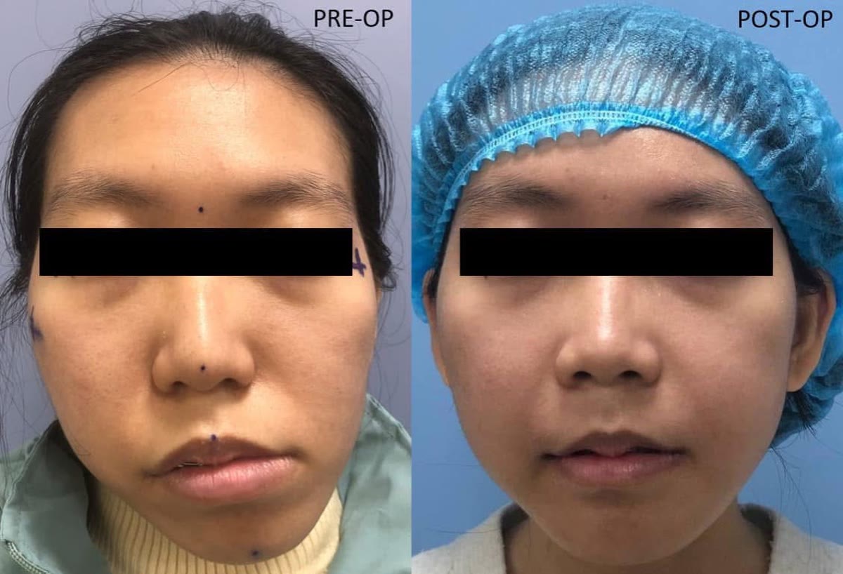
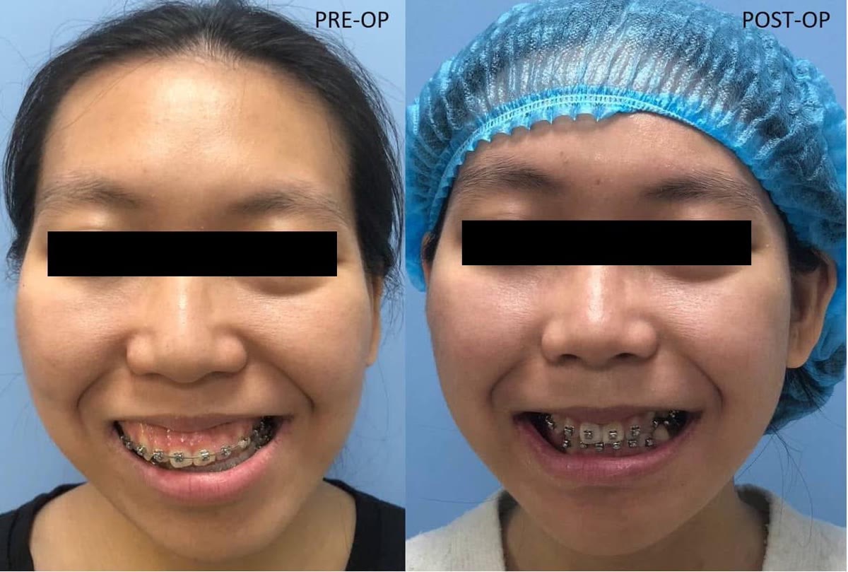
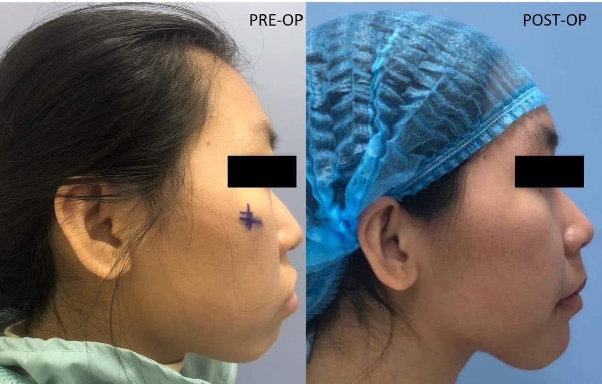
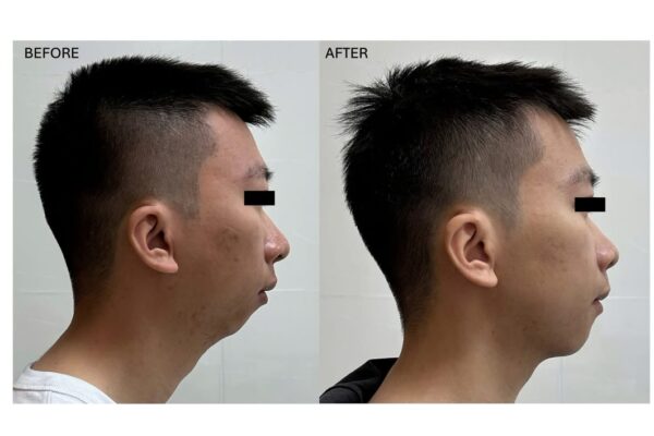
Đây là một trong những trường hợp phẫu thuật chỉnh hình xương hàm phức tạp mà mình cùng ê-kíp đã thực hiện, với sự kết hợp của ba vấn đề: hô hai hàm, cười hở lợi và bất đối xứng khuôn mặt.
Bệnh nhân là du học sinh và chỉ có một khoảng thời gian tương đối ngắn tại Việt Nam. Trước đó, bệnh nhân đã được chỉnh nha trong vài năm để chuẩn bị cho phẫu thuật.
Qua thăm khám và phân tích, bệnh nhân được chẩn đoán quá phát hai hàm (bimaxillary protrusion), quá phát hàm trên theo chiều dọc (vertical maxillary excess) gây cười hở lợi, đồng thời có bất đối xứng xương hàm và lệch mặt.
Việc điều trị vì vậy không chỉ đơn thuần là đưa hai hàm ra sau, mà phải đồng thời kiểm soát vị trí của hàm trên theo ba chiều, độ nhô của vùng răng cửa, tương quan khớp cắn, bất đối xứng và hình thái mô mềm của khuôn mặt.
Kế hoạch phẫu thuật
Ở hàm trên, Le Fort I osteotomy được thực hiện nhằm đưa hàm trên lên trên, qua đó điều chỉnh tình trạng quá phát theo chiều dọc và giảm mức độ cười hở lợi.
Tuy nhiên, bệnh nhân đồng thời có tình trạng nhô đáng kể của vùng răng cửa. Vì vậy, anterior segmental osteotomy (ASO) được kết hợp để đưa phân đoạn răng trước hàm trên ra sau.
Việc phân chia chuyển động theo cách này cho phép kiểm soát tương đối độc lập hai thành phần của biến dạng: vị trí theo chiều dọc của toàn bộ hàm trên và độ nhô trước – sau của phân đoạn răng cửa, thay vì phải lùi toàn bộ hàm trên một khoảng lớn.
Ở hàm dưới, bilateral sagittal split osteotomy (BSSO) được sử dụng để điều chỉnh vị trí và bất đối xứng của xương hàm dưới. ASO hàm dưới được thực hiện đồng thời nhằm đưa phân đoạn răng cửa dưới ra sau và cải thiện tình trạng hô hai hàm.
Khi xương cân nhưng khuôn mặt vẫn chưa cân
Một trong những phần khó nhất của trường hợp này là genioplasty – phẫu thuật tạo hình cằm.
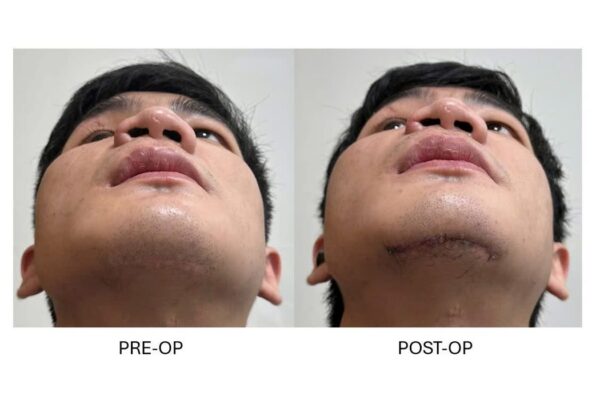
Nếu chỉ đưa mảnh cằm về vị trí đối xứng dựa trên các mốc xương, mô mềm vùng cằm vẫn biểu hiện lệch sang trái. Đây là một vấn đề quan trọng trong phẫu thuật chỉnh hình xương hàm: đối xứng của bộ xương không phải lúc nào cũng tạo ra đối xứng của mô mềm.
Khuôn mặt cuối cùng mà chúng ta quan sát là mô mềm, không phải hình ảnh của bộ xương trên CT.
Vì vậy, trong trường hợp này, mảnh cằm được chủ động dịch sang phải nhiều hơn so với vị trí đối xứng lý tưởng của xương – một dạng overcorrection có chủ đích. Điều này đồng nghĩa với việc chấp nhận một mức độ bất đối xứng nhất định ở cấu trúc xương để đạt được sự cân bằng tốt hơn của khuôn mặt nhìn từ bên ngoài.
Đây cũng là một ví dụ cho thấy lập kế hoạch phẫu thuật chỉnh hình xương hàm không thể chỉ dựa trên các con số và đường tham chiếu của bộ xương. Mục tiêu cuối cùng là sự hài hòa của khuôn mặt, chức năng và khớp cắn.
Kết quả
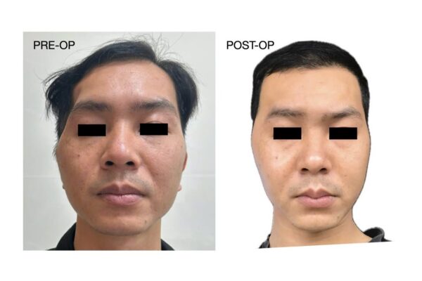
Sau phẫu thuật, tình trạng hô hai hàm, cười hở lợi và bất đối xứng khuôn mặt đều được cải thiện đáng kể. Khớp cắn đạt được tương quan lồng múi tương đối tốt và chức năng ăn nhai gần như bình thường.
Như trong mọi phẫu thuật chỉnh hình xương hàm, kết quả thực tế có thể có những sai khác nhỏ so với kế hoạch phẫu thuật ảo. Điều quan trọng là đánh giá kết quả tổng thể dựa trên sự cân đối của khuôn mặt, tương quan mô mềm, khớp cắn và chức năng, thay vì chỉ dựa vào mức độ trùng khớp tuyệt đối giữa kế hoạch và vị trí xương sau phẫu thuật.
Quan trọng hơn, sự thay đổi về khuôn mặt đã giúp bệnh nhân cảm thấy tự tin hơn rất nhiều.
Sau phẫu thuật, bệnh nhân tiếp tục giai đoạn chỉnh nha hoàn thiện nhằm tinh chỉnh khớp cắn và duy trì kết quả điều trị lâu dài.
This was one of the more challenging orthognathic cases managed by our team, combining three distinct dentofacial problems: bimaxillary protrusion, excessive gingival display, and facial asymmetry.
The patient was studying abroad and had only a limited period of time available in Vietnam. She had previously undergone several years of orthodontic preparation for orthognathic surgery.
Clinical and radiographic assessment demonstrated bimaxillary protrusion, vertical maxillary excess, resulting in excessive gingival display on smiling, together with skeletal asymmetry.
The surgical objective was therefore not simply to move both jaws posteriorly. The treatment had to simultaneously address the three-dimensional position of the maxilla, anterior dental protrusion, occlusion, skeletal asymmetry, and the resulting soft-tissue appearance.
Surgical planning
In the maxilla, a Le Fort I osteotomy was performed with superior repositioning to correct the vertical maxillary excess and reduce excessive gingival display.
However, the patient also presented with significant protrusion of the anterior dentoalveolar segment. An anterior segmental osteotomy (ASO) was therefore incorporated to allow posterior repositioning of the maxillary anterior segment.
This strategy allowed two components of the deformity to be controlled relatively independently: the vertical position of the maxilla as a whole and the anteroposterior position of the anterior dentoalveolar segment, avoiding the need for excessive posterior repositioning of the entire maxilla.
In the mandible, a bilateral sagittal split osteotomy (BSSO) was performed to correct mandibular position and asymmetry. A mandibular ASO was simultaneously performed to retract the lower anterior dentoalveolar segment and further correct the bimaxillary protrusion.
When skeletal symmetry does not produce facial symmetry
One of the most challenging components of this case was the genioplasty.
When the chin segment was positioned according to skeletal symmetry alone, the overlying soft-tissue chin remained visibly deviated to the left.
This illustrates an important consideration in orthognathic surgery: skeletal symmetry does not necessarily translate directly into soft-tissue symmetry.
Ultimately, what determines the visible facial result is the soft-tissue envelope rather than the skeletal anatomy seen on CT.
For this reason, the chin segment was intentionally translated further toward the right than would have been required to achieve ideal skeletal symmetry. In other words, a degree of planned overcorrection was accepted at the skeletal level in order to achieve a more balanced external facial appearance.
This case demonstrates why orthognathic surgical planning cannot rely exclusively on skeletal landmarks, reference lines, or numerical measurements. The ultimate objective is harmony between facial appearance, soft-tissue balance, function, and occlusion.
Outcome
Following surgery, substantial improvement was achieved in the patient's bimaxillary protrusion, excessive gingival display, and facial asymmetry. A reasonably good intercuspation was established, and masticatory function returned to near normal.
As with any orthognathic procedure, minor discrepancies may exist between virtual surgical planning and the final postoperative result. The overall outcome should therefore be evaluated not simply by absolute correspondence between the planned and postoperative skeletal positions, but by considering facial balance, the soft-tissue response, occlusion, and function as a whole.
Most importantly, the improvement in facial balance resulted in a significant increase in the patient's confidence.
Postoperative orthodontic treatment was subsequently continued to refine the occlusion and consolidate the final result.