Phẫu thuật le fort ii kết hợp le fort i và bsso điều trị sai khớp cắn loại iii trên bệnh nhân khe hở môi – vòm miệngCombined Le Fort ii, Le Fort i, and BSSO for cleft-related skeletal class iii deformity
02/09/2026Kết quảResults
Le Fort I là một trong những kỹ thuật phổ biến nhất trong phẫu thuật chỉnh hình xương hàm. Đường cắt này cho phép di chuyển phần xương hàm trên mang răng theo ba chiều không gian, nhờ đó có thể điều chỉnh nhiều dạng sai lệch xương hàm và khớp cắn.
Tuy nhiên, ở một số bệnh nhân, đặc biệt là những trường hợp có dị tật bẩm sinh, sự kém phát triển không chỉ giới hạn ở phần xương hàm trên mang răng mà còn liên quan đến toàn bộ vùng mũi – hàm trên.
Trong những trường hợp như vậy, Le Fort I đơn thuần có thể không đủ để giải quyết toàn bộ biến dạng khuôn mặt.
Kém phát triển tầng mặt giữa trên bệnh nhân khe hở môi – vòm miệng
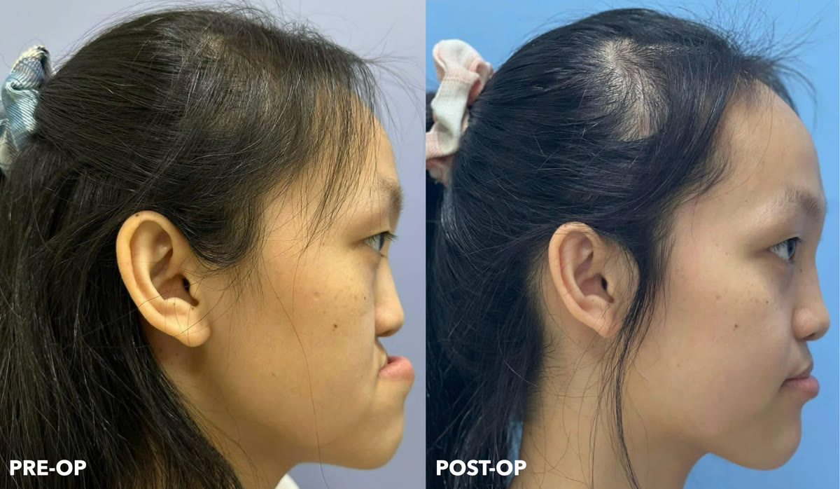
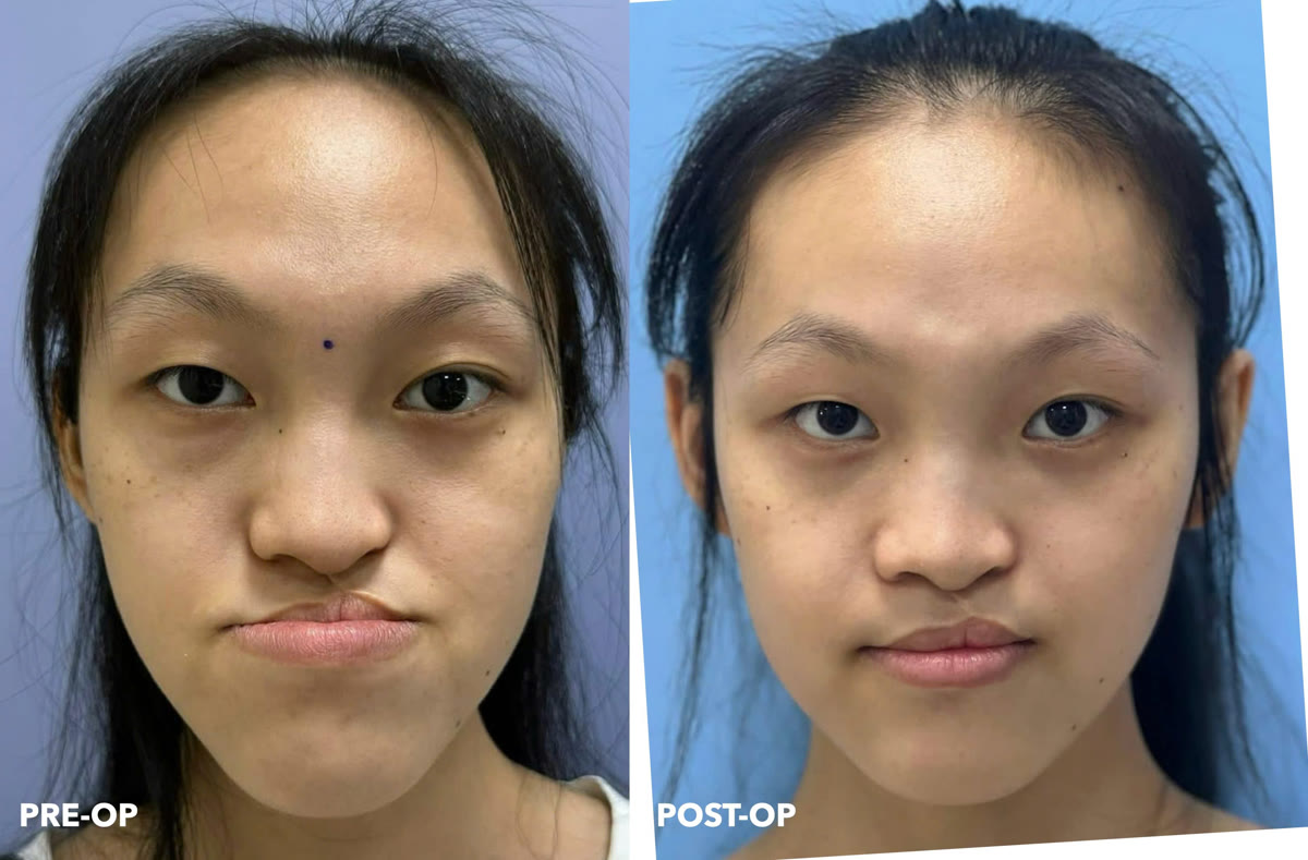
Bệnh nhân nữ, 20 tuổi, có tiền sử khe hở môi – vòm miệng và biểu hiện sai khớp cắn loại III do xương kèm kém phát triển nặng tầng mặt giữa.
Qua đánh giá khuôn mặt và hình ảnh ba chiều, tình trạng thiếu phát triển tập trung chủ yếu ở phức hợp mũi – hàm trên, trong khi vùng gò má và ổ mắt tương đối bình thường.
Đây là yếu tố quan trọng quyết định lựa chọn đường cắt xương.
Nếu chỉ thực hiện Le Fort I và đưa hàm trên ra trước, phần xương mang răng có thể đạt được tương quan phù hợp với hàm dưới. Tuy nhiên, phần mũi và vùng giữa mặt phía trên đường cắt sẽ không được di chuyển tương ứng.
Kết quả có thể là khớp cắn được cải thiện nhưng sự thiếu hụt của vùng mũi – tầng mặt giữa vẫn còn, thậm chí sự không tương xứng giữa vùng mũi và phần hàm trên đã được đưa ra trước có thể trở nên rõ hơn.
Vì vậy, mục tiêu của phẫu thuật trong trường hợp này không chỉ là điều chỉnh khớp cắn mà còn phải tái lập sự cân đối của toàn bộ tầng mặt giữa.
Tại sao lựa chọn Le Fort II?
Khác với Le Fort I, đường cắt Le Fort II nằm ở vị trí cao hơn và cho phép di chuyển đồng thời phức hợp mũi – hàm trên.
Do vùng gò má và ổ mắt của bệnh nhân tương đối bình thường, việc lựa chọn Le Fort II cho phép tập trung điều chỉnh chính xác vùng bị thiếu phát triển mà không cần di chuyển toàn bộ tầng mặt giữa ở mức cao hơn.
Sau khi thảo luận kế hoạch điều trị cùng Giáo sư Imad Abu El-Naaj, mình quyết định thực hiện phối hợp ba đường cắt xương:
Le Fort II để đưa toàn bộ phức hợp mũi – hàm trên ra trước.
Le Fort I để tiếp tục tinh chỉnh vị trí của phần hàm trên mang răng, cho phép kiểm soát tốt hơn tương quan xương và khớp cắn.
BSSO hai bên ở hàm dưới để đưa xương hàm dưới về vị trí phù hợp với vị trí mới của hàm trên và thiết lập tương quan khớp cắn mong muốn.
Việc phối hợp Le Fort II và Le Fort I cho phép hai thành phần của tầng mặt giữa được điều chỉnh ở những mức độ khác nhau, thay vì buộc toàn bộ cấu trúc phải di chuyển như một khối duy nhất.
Lập kế hoạch phẫu thuật trên môi trường số
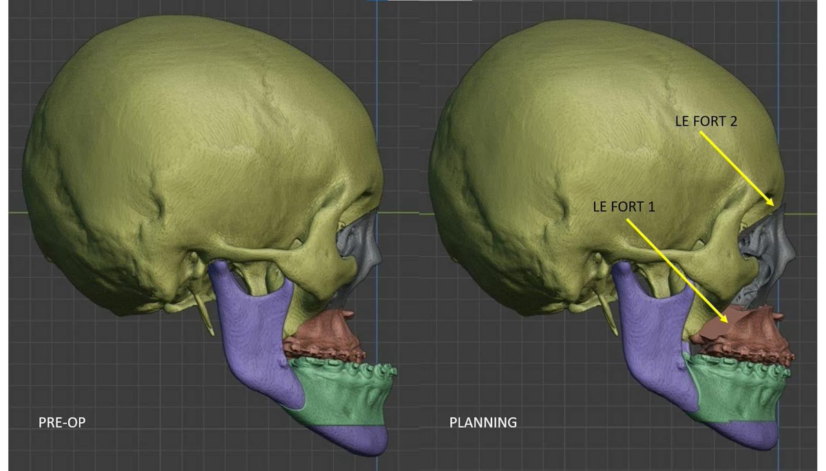
Do sự phức tạp của các đường cắt và chuyển động xương, toàn bộ ca phẫu thuật được lập kế hoạch trước trên mô hình ba chiều.
Dựa trên dữ liệu hình ảnh của bệnh nhân, các đường cắt xương và vị trí dự kiến của từng phân đoạn sau phẫu thuật được mô phỏng trên máy tính. Nhờ đó, có thể đánh giá trước tương quan giữa vùng mũi, hàm trên, hàm dưới và khớp cắn trước khi thực hiện phẫu thuật thực tế.
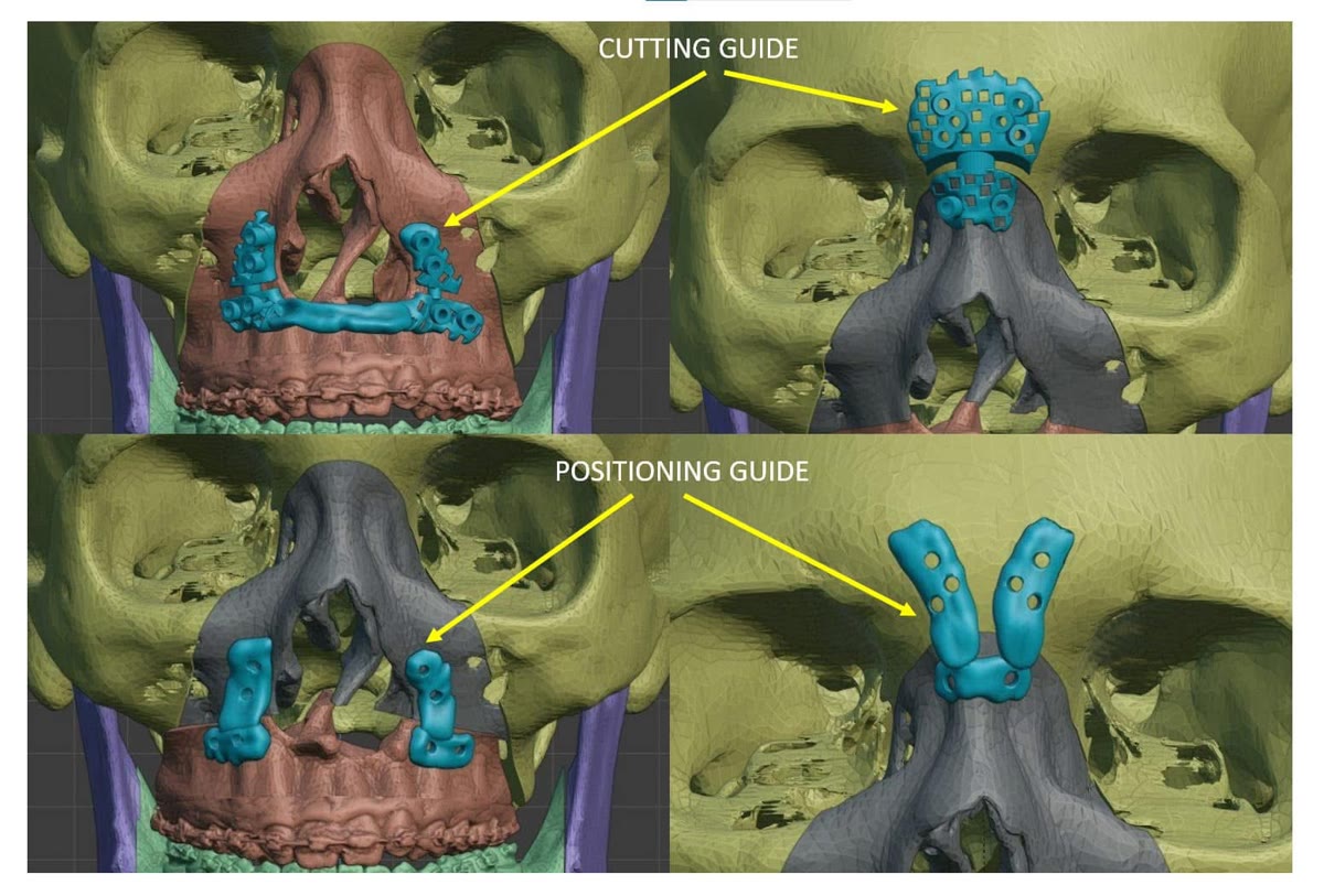
Sau khi hoàn thiện kế hoạch, máng hướng dẫn cắt xương và dụng cụ định vị xương cá nhân hóa được thiết kế dựa trên giải phẫu riêng của bệnh nhân và chế tạo bằng công nghệ in 3D kim loại.
Máng hướng dẫn cắt giúp chuyển vị trí các đường cắt đã thiết kế trên máy tính vào phẫu trường.
Sau khi xương được di chuyển, dụng cụ định vị cá nhân hóa được sử dụng để hướng dẫn các phân đoạn xương đến vị trí đã xác định trong kế hoạch.
Trong kỹ thuật được sử dụng ở trường hợp này, dụng cụ định vị chỉ đóng vai trò tạm thời trong quá trình xác lập vị trí xương. Sau khi vị trí mong muốn được xác nhận, dụng cụ được tháo bỏ và các phân đoạn xương được cố định bằng hệ thống nẹp vít thương mại thông thường.
Như vậy, công nghệ cá nhân hóa được sử dụng để chuyển kế hoạch số vào phẫu thuật, nhưng không trở thành vật liệu cố định vĩnh viễn của bệnh nhân.
Điều trị không chỉ nhằm tạo ra một khớp cắn tốt
Trên những bệnh nhân có biến dạng sọ mặt bẩm sinh, mục tiêu của phẫu thuật không thể chỉ giới hạn ở việc đưa răng về tương quan khớp cắn loại I.
Một khớp cắn tốt nhưng tầng mặt giữa vẫn thiếu phát triển chưa phải là một kết quả hoàn chỉnh.
Việc lựa chọn mức cắt xương cần dựa trên vị trí thực sự của biến dạng trên khuôn mặt: nếu vấn đề nằm ở phần xương ổ răng và hàm trên, Le Fort I có thể phù hợp; nếu sự thiếu phát triển kéo dài lên vùng mũi – hàm trên, một đường cắt cao hơn như Le Fort II có thể cần được cân nhắc.
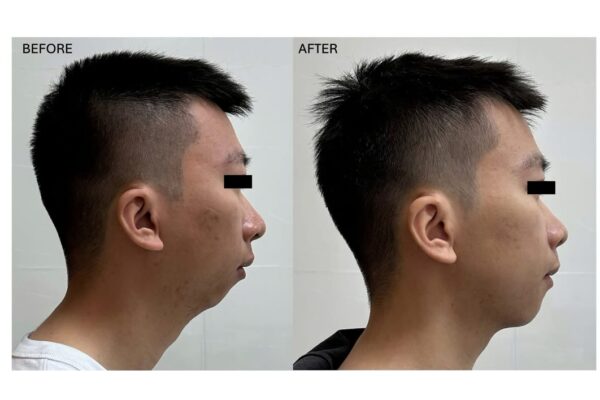
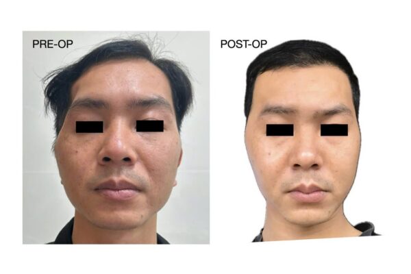
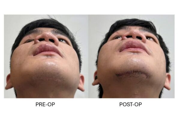
Sau phẫu thuật, tương quan hai hàm và hình thái tầng mặt giữa của bệnh nhân đã được cải thiện đáng kể. Bệnh nhân và gia đình hài lòng với kết quả đạt được.
Do đây là một trường hợp dị tật sọ mặt bẩm sinh phức tạp, phẫu thuật chỉnh hình xương hàm chỉ là một phần trong toàn bộ quá trình điều trị. Bệnh nhân vẫn có thể cần những can thiệp tiếp theo để hoàn thiện thêm hình thái và chức năng của khuôn mặt.
Đây cũng là trường hợp Le Fort II đầu tiên được thực hiện tại Bệnh viện Răng Hàm Mặt Trung ương Hà Nội theo ghi nhận của ê-kíp tại thời điểm phẫu thuật, đồng thời là một ca bệnh đặc biệt với sự phối hợp Le Fort II, Le Fort I và BSSO dưới sự hỗ trợ của lập kế hoạch phẫu thuật ba chiều và các dụng cụ phẫu thuật cá nhân hóa.
Le Fort I osteotomy is one of the most commonly performed procedures in orthognathic surgery. It allows the tooth-bearing maxilla to be repositioned in three dimensions and can therefore address a wide range of skeletal and occlusal discrepancies.
In some patients, however, particularly those with congenital craniofacial deformities, the deficiency is not confined to the tooth-bearing maxilla. Instead, hypoplasia may involve the entire nasomaxillary complex.
In such cases, a conventional Le Fort I advancement alone may not adequately address the facial deformity.
Midfacial deficiency in a patient with cleft lip and palate
The patient was a 20-year-old woman with a history of cleft lip and palate who presented with a severe skeletal Class III deformity associated with significant midfacial hypoplasia.
Clinical and three-dimensional assessment demonstrated that the deficiency predominantly involved the nasomaxillary complex, while the zygomatic and orbital regions were relatively well developed.
This distribution of the deformity was a critical factor in determining the appropriate level of osteotomy.
If a conventional Le Fort I advancement had been performed alone, the tooth-bearing maxilla could have been advanced into an appropriate relationship with the mandible. However, the nasal complex and the midface above the Le Fort I level would have remained relatively retruded.
The result could therefore have been an improved occlusion without adequate correction of the midfacial deficiency, potentially making the discrepancy between the advanced maxilla and the retruded nasal region even more apparent.
The objective of surgery was therefore not simply to correct the bite, but to restore a more harmonious relationship of the entire midface.
Why Le Fort II?
Unlike a Le Fort I osteotomy, a Le Fort II osteotomy is positioned at a higher level and allows the nasomaxillary complex to be advanced as a unit.
Because the patient's zygomatic and orbital regions were relatively well developed, a Le Fort II osteotomy allowed the operation to specifically address the deficient nasomaxillary region without unnecessarily mobilizing the entire upper midface.
Following discussion of the surgical strategy with Professor Imad Abu El-Naaj, I elected to combine three osteotomies:
A Le Fort II osteotomy was used to advance the nasomaxillary complex.
A Le Fort I osteotomy was incorporated to provide additional control of the tooth-bearing maxilla, allowing further refinement of the skeletal and occlusal relationship.
A bilateral sagittal split osteotomy (BSSO) was performed in the mandible to reposition the lower jaw according to the new maxillary position and establish the intended occlusion.
The combination of Le Fort II and Le Fort I allowed different components of the midface to be repositioned by different amounts rather than requiring the entire region to move as a single unit.
Three-dimensional surgical planning
Because of the complexity of the osteotomies and skeletal movements, the entire procedure was planned preoperatively in a three-dimensional virtual environment.
Using the patient's imaging data, the osteotomy lines and intended postoperative positions of the individual skeletal segments were simulated before surgery. This allowed the relationship between the nasal complex, maxilla, mandible, and occlusion to be evaluated before transferring the plan to the operating room.
Once the surgical plan had been finalized, patient-specific cutting guides and temporary positioning guides were designed according to the patient's individual anatomy and manufactured using metal 3D printing.
The cutting guides were used to transfer the planned osteotomy lines accurately to the surgical field.
Following mobilization of the skeletal segments, the patient-specific positioning guides were used to reproduce the planned postoperative skeletal positions.
Importantly, in this technique the patient-specific positioning devices served only as temporary intraoperative guides. Once the intended skeletal position had been established and verified, they were removed and replaced with conventional commercially available fixation plates and screws.
In this way, patient-specific technology was used to transfer the virtual surgical plan to the operating room without requiring permanent patient-specific fixation implants.
Beyond correction of the occlusion
In patients with congenital craniofacial deformities, successful orthognathic surgery cannot be defined solely by achieving a Class I dental relationship.
A satisfactory occlusion with persistent midfacial deficiency does not represent complete correction of the deformity.
The level of osteotomy should therefore be selected according to where the skeletal deficiency actually exists within the face. When the deformity is primarily dentoalveolar or maxillary, a Le Fort I osteotomy may be sufficient. When the deficiency extends superiorly into the nasomaxillary complex, a higher-level osteotomy such as Le Fort II may need to be considered.
Following surgery, the patient's maxillomandibular relationship and midfacial projection were substantially improved, and both the patient and her family were satisfied with the outcome.
Because this was a complex congenital craniofacial deformity, orthognathic surgery represented one component of a longer reconstructive pathway. Additional procedures may still be required to further refine facial form and function.
According to our team's records at the time of surgery, this was the first Le Fort II osteotomy performed at the National Hospital of Odonto-Stomatology, Hanoi. It was also a particularly challenging case combining Le Fort II, Le Fort I, and BSSO with three-dimensional virtual surgical planning and patient-specific surgical guides.