Điều trị sai khớp cắn loại ii kết hợp hô xương ổ răng hai hàmManagement of class ii malocclusion with bimaxillary dentoalveolar protrusion
02/09/2026Kết quảResults
Hô vùng miệng không phải lúc nào cũng đơn thuần do vị trí của xương hàm. Ở một số bệnh nhân, tình trạng này là sự kết hợp giữa sai lệch vị trí của nền xương hàm và sự nhô ra trước của xương ổ răng cùng các răng cửa.
Trường hợp này là một ví dụ điển hình: bệnh nhân có sai khớp cắn loại II với hàm dưới và cằm lùi, đồng thời có hô xương ổ răng hai hàm. Việc điều trị vì vậy cần giải quyết đồng thời cả hai thành phần để cải thiện khớp cắn và hình thái khuôn mặt.
Từ một trường hợp chỉnh nha kéo dài đến chỉ định phẫu thuật
Bệnh nhân đã trải qua nhiều năm điều trị chỉnh nha nhưng tình trạng khuôn mặt và tương quan xương hàm không được cải thiện như mong muốn. Sau đó, bệnh nhân được một đồng nghiệp chỉnh nha giới thiệu đến mình để đánh giá khả năng điều trị bằng phẫu thuật chỉnh hình xương hàm.
Khám lâm sàng kết hợp với phân tích hình ảnh cho thấy một số đặc điểm chính:
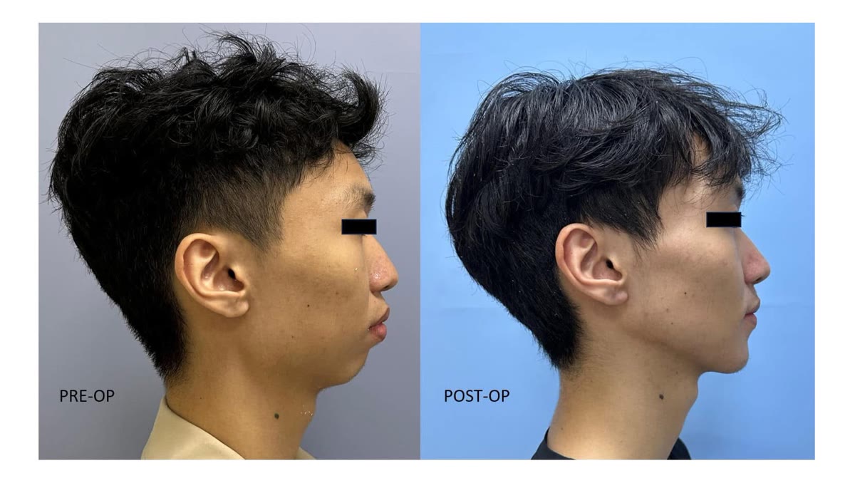
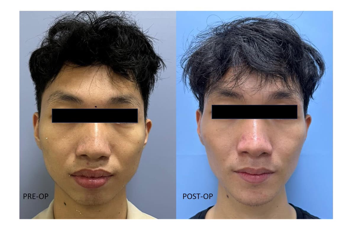
Vùng môi và xương ổ răng nhô ra trước, góc mũi – môi nhọn.
Hàm dưới lùi ra sau so với tầng mặt giữa.
Cằm lùi, làm tăng cảm giác lồi của khuôn mặt khi nhìn nghiêng.
Các răng cửa dưới nghiêng nhiều ra trước, đi kèm với sự nhô của phân đoạn xương ổ răng phía trước.
Bệnh nhân đồng thời mong muốn cải thiện độ rộng và hình thể vùng góc hàm.
Vì vậy, nếu chỉ đưa hàm dưới ra trước hoặc chỉ kéo lùi các răng cửa sẽ không giải quyết đầy đủ toàn bộ biến dạng.
Kế hoạch phẫu thuật
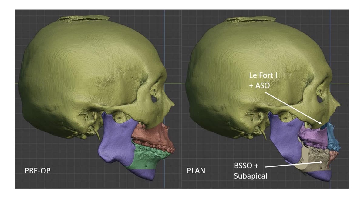
Ở hàm trên, răng 14 và 24 được nhổ để tạo khoảng. Sau đó thực hiện Le Fort I và chia hàm trên thành ba phân đoạn qua vùng nhổ răng.
Kỹ thuật này cho phép kiểm soát riêng phân đoạn răng trước, vừa điều chỉnh lại trục của các răng cửa trên vừa đưa toàn bộ phân đoạn này ra sau. Mục tiêu là giảm độ nhô của vùng hàm trên và cải thiện tương quan giữa mũi, môi và tầng mặt dưới.
Ở hàm dưới, răng 34 và 44 được nhổ. Phẫu thuật chẻ dọc cành cao hai bên được kết hợp với đường cắt xương dưới chóp các răng cửa.
Hai chuyển động khác nhau được thực hiện đồng thời: phần hàm dưới phía sau được đưa ra trước để điều chỉnh tình trạng lùi hàm, trong khi phân đoạn răng cửa được kiểm soát riêng để dựng lại trục răng và giảm độ nhô của vùng xương ổ răng phía trước.
Nhờ đó, có thể đưa nền xương hàm dưới ra trước nhưng đồng thời kiểm soát vị trí của các răng cửa, thay vì buộc hai thành phần này phải di chuyển theo cùng một hướng.
Cuối cùng, vùng góc hàm hai bên được tạo hình nhằm cải thiện đường viền tầng mặt dưới theo mong muốn thẩm mỹ của bệnh nhân.
Điều trị khuôn mặt chứ không chỉ điều trị vị trí của răng
Điểm quan trọng của trường hợp này là sự khác biệt giữa vị trí của nền xương hàm và vị trí của xương ổ răng cùng các răng trên nền xương đó.
Một bệnh nhân có thể đồng thời có hàm dưới lùi nhưng các răng cửa dưới lại nghiêng và nhô ra trước. Nếu chỉ nhìn vào vị trí của răng, việc đưa hàm dưới ra trước có vẻ mâu thuẫn với mục tiêu giảm hô. Nhưng khi phân tích riêng từng thành phần, hai vấn đề hoàn toàn có thể được điều chỉnh đồng thời.
Đây cũng là lý do phẫu thuật phân đoạn xương ổ răng được kết hợp với phẫu thuật chỉnh hình hai hàm trong trường hợp này.
Lập kế hoạch và chuyển kế hoạch vào phẫu thuật
Toàn bộ các chuyển động xương được lập kế hoạch trước trên môi trường ba chiều.
Việc mô phỏng trước phẫu thuật cho phép đánh giá đồng thời vị trí của hai hàm, các phân đoạn xương ổ răng, trục răng cửa, cằm, khớp cắn và hình thái khuôn mặt trước khi quyết định phương án cuối cùng.
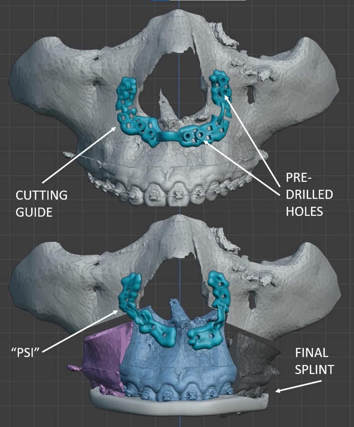
Sau khi hoàn thiện kế hoạch, các máng hướng dẫn và dụng cụ định vị cá nhân hóa được thiết kế riêng cho bệnh nhân nhằm hỗ trợ chuyển vị trí xương đã xác định trên máy tính vào phẫu trường.
Việc lập kế hoạch kỹ thuật số và sử dụng các dụng cụ hướng dẫn cá nhân hóa đã trở thành một phần thường quy trong các ca phẫu thuật chỉnh hình xương hàm do mình thực hiện từ năm 2021.
Vai trò của chỉnh nha sau phẫu thuật
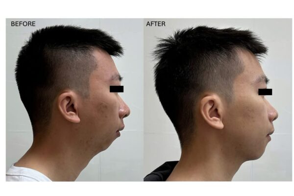
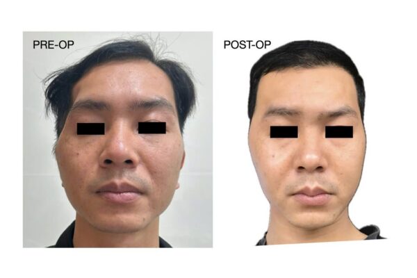
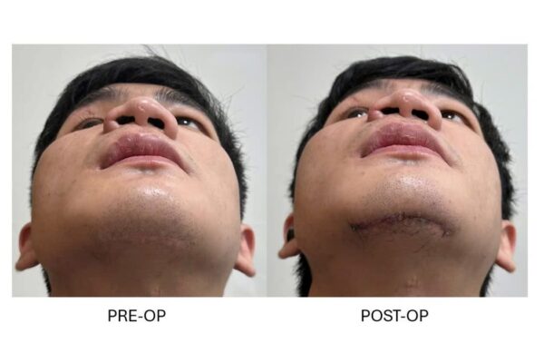
Khoảng năm tháng sau phẫu thuật, bệnh nhân có sự cải thiện rõ rệt về tương quan khuôn mặt và khớp cắn. Độ nhô vùng miệng giảm, hàm dưới và cằm được đưa ra trước, tạo sự cân bằng tốt hơn giữa các tầng mặt. Chức năng ăn nhai, há miệng và phát âm ổn định.
Tuy nhiên, phẫu thuật không phải là bước cuối cùng của quá trình điều trị.
Chỉnh nha sau phẫu thuật đóng vai trò quan trọng trong việc tinh chỉnh vị trí từng răng, hoàn thiện sự lồng múi và ổn định khớp cắn. Kết quả của một ca phẫu thuật chỉnh hình xương hàm vì vậy không chỉ phụ thuộc vào phẫu thuật viên mà còn cần sự phối hợp chặt chẽ giữa phẫu thuật và chỉnh nha trong toàn bộ quá trình điều trị.
Trường hợp này cho thấy một nguyên tắc quan trọng: để điều trị hô vùng miệng hiệu quả, trước hết cần xác định chính xác phần nào của biến dạng đến từ nền xương hàm và phần nào đến từ xương ổ răng cùng vị trí của các răng.
Protrusion of the lower face is not always caused simply by the position of the jaws. In some patients, the deformity results from a combination of skeletal jaw discrepancy and protrusion of the anterior dentoalveolar segments.
This case illustrates such a combination: a skeletal Class II pattern with mandibular and chin retrusion, together with bimaxillary dentoalveolar protrusion. Successful treatment therefore required independent correction of both components to improve the facial profile and occlusion.
From prolonged orthodontic treatment to orthognathic surgery
The patient had undergone orthodontic treatment for several years without achieving the desired correction of the underlying facial and skeletal discrepancy. He was subsequently referred by an orthodontic colleague for assessment for orthognathic surgery.
Clinical and radiographic evaluation demonstrated several key features:
Prominence of the lips and anterior dentoalveolar region with an acute nasolabial angle.
Mandibular retrusion relative to the midface.
A retruded chin contributing to a convex facial profile.
Significant proclination of the lower incisors associated with anterior dentoalveolar protrusion.
A desire for additional refinement of the mandibular angle and lower facial contour.
Simply advancing the mandible or retracting the anterior teeth alone would therefore have been insufficient to address the complete deformity.
Surgical strategy
In the maxilla, the first premolars were extracted to create the space required for segmental repositioning. A Le Fort I osteotomy was then performed and the maxilla divided into three segments through the extraction sites.
This allowed independent control of the anterior maxillary segment, with correction of upper incisor inclination and posterior repositioning of the anterior dentoalveolar complex. The objective was to reduce maxillary protrusion and improve the relationship between the nose, lips, and lower facial profile.
In the mandible, the first premolars were also extracted. A bilateral sagittal split osteotomy was combined with an anterior subapical osteotomy.
This enabled two different movements to be performed simultaneously: the mandibular basal segment could be advanced to correct mandibular retrusion, while the anterior dentoalveolar segment could be independently repositioned to upright the lower incisors and reduce anterior protrusion.
In other words, the operation allowed us to advance the mandible while independently controlling the position of the lower incisors, rather than forcing the skeletal base and dentoalveolar segment to move together.
Bilateral mandibular angle contouring was additionally performed to refine the lower facial contour according to the patient’s aesthetic objectives.
Treating the face, not simply the teeth
An important aspect of this case is the distinction between the position of the skeletal jaw base and the position of the dentoalveolar structures supported by that base.
A patient may simultaneously have a retruded mandible and markedly proclined, protrusive lower incisors. When the teeth are considered in isolation, mandibular advancement may appear contradictory to the objective of reducing protrusion. Once the skeletal and dentoalveolar components are analyzed separately, however, both problems can be corrected simultaneously.
This was the rationale for combining segmental dentoalveolar osteotomies with bimaxillary orthognathic surgery in this patient.
Digital planning and surgical transfer
All skeletal movements were planned preoperatively in a three-dimensional virtual environment.
Virtual planning allowed the positions of the jaws, dentoalveolar segments, incisor inclinations, chin, occlusion, and overall facial profile to be assessed together before the final surgical movements were determined.
Once the plan had been finalized, patient-specific surgical guides and positioning devices were designed to assist in transferring the planned skeletal positions from the virtual environment to the operating room.
Digital planning and individualized surgical guidance have been incorporated routinely into my orthognathic surgery workflow since 2021.
The role of postoperative orthodontics
At approximately five months after surgery, the patient demonstrated substantial improvement in facial balance and occlusal relationship. The perioral protrusion was reduced, while advancement of the mandible and chin created a more balanced facial profile. Mastication, mouth opening, and speech were functioning well.
Surgery, however, does not represent the end of treatment.
Postoperative orthodontics plays an essential role in refining individual tooth positions, improving intercuspation, and consolidating the final occlusion. A successful orthognathic outcome therefore depends on close coordination between the surgeon and orthodontist throughout the treatment process.
This case illustrates an important principle: effective correction of facial protrusion begins by determining how much of the deformity originates from the skeletal jaw bases and how much originates from the dentoalveolar segments and tooth position.