Kéo giãn sinh xương điều trị thiểu sản hàm dưới nặng kèm thiếu lồi cầu bẩm sinhMandibular distraCTion osteogenesis for severe hypoplasia with congenital absence of the condyle
02/09/2026Kết quảResults
Điều trị biến dạng xương hàm ở trẻ em luôn đặt ra một câu hỏi khó: nên can thiệp sớm hay chờ đến khi bệnh nhân hoàn tất tăng trưởng?
Đối với nhiều biến dạng xương hàm, phẫu thuật chỉnh hình xương hàm toàn diện thường được trì hoãn đến khi quá trình tăng trưởng gần hoàn tất. Tuy nhiên, khi biến dạng quá nặng và đã ảnh hưởng đến những chức năng quan trọng như hô hấp, ăn nhai và sự phát triển của hệ thống sọ mặt, việc chờ đợi không phải lúc nào cũng là lựa chọn phù hợp.
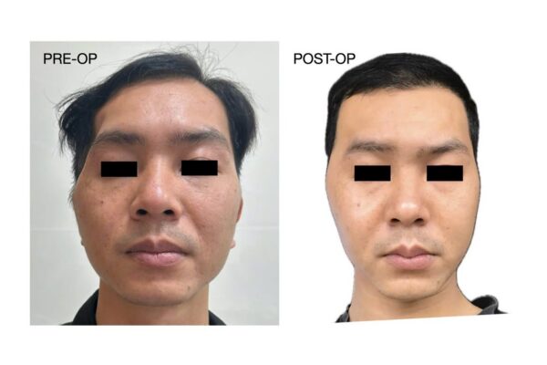
Trường hợp này là một bệnh nhân 11 tuổi với thiểu sản xương hàm dưới rất nặng, bất đối xứng sọ mặt và thiếu lồi cầu xương hàm dưới phải bẩm sinh.
Thiểu sản hàm dưới không chỉ là vấn đề thẩm mỹ
Bệnh nhân có khe hở ngang mặt Tessier số 7 kèm teo nửa mặt phải. Xương hàm dưới bên phải thiểu sản nghiêm trọng và không có lồi cầu bẩm sinh.
Toàn bộ xương hàm dưới có kích thước nhỏ và lùi nhiều ra sau. Tình trạng này không chỉ tạo ra biến dạng khuôn mặt mà còn liên quan đến nhiều vấn đề chức năng.
Các răng chen chúc do cung hàm kém phát triển. Quan trọng hơn, vị trí lùi của hàm dưới góp phần làm giảm không gian đường thở phía sau lưỡi và bệnh nhân được xác định có ngưng thở khi ngủ tắc nghẽn.
Bệnh nhân từng được thăm khám nhưng việc điều trị gặp khó khăn do bệnh nhân vẫn đang trong giai đoạn tăng trưởng.
Tuy nhiên, trong trường hợp này, mục tiêu của can thiệp sớm không phải là tạo ra kết quả khuôn mặt cuối cùng ở tuổi 11. Mục tiêu trước mắt là cải thiện cấu trúc hàm dưới, chức năng hô hấp và ăn nhai, đồng thời tạo điều kiện thuận lợi hơn cho quá trình phát triển và các bước điều trị tiếp theo.
Một kế hoạch điều trị theo nhiều giai đoạn
Sau khi hội chẩn với TS. Sang Nguyen Minh về tình trạng hô hấp khi ngủ và trao đổi kế hoạch điều trị dưới sự hướng dẫn của Giáo sư Imad Abu El-Naaj, phương án được xây dựng theo nhiều giai đoạn.
Giai đoạn đầu: tái tạo lồi cầu
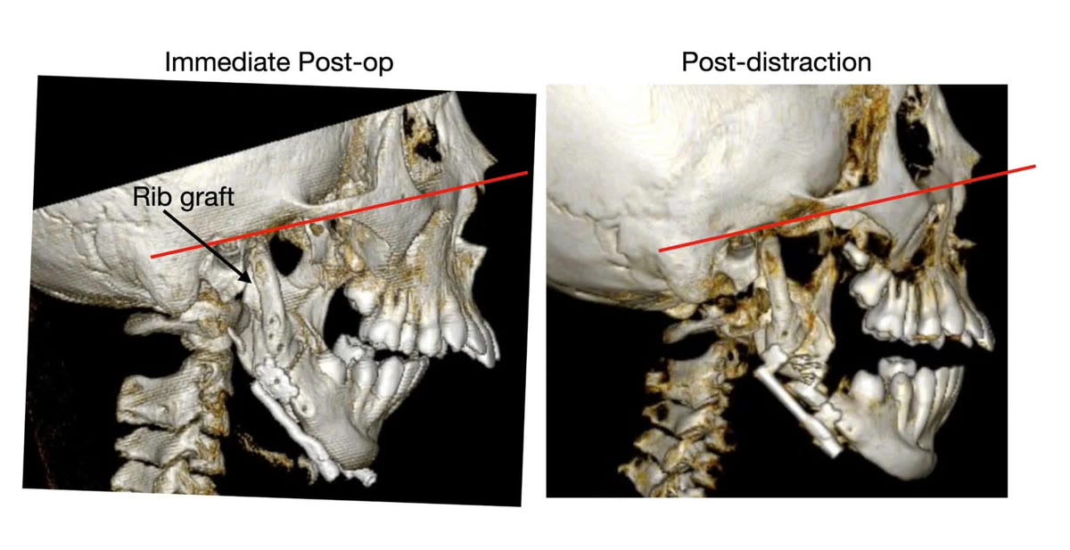
Do bệnh nhân thiếu lồi cầu xương hàm dưới phải bẩm sinh, bước đầu tiên là tái tạo vùng lồi cầu bằng mảnh ghép sụn – sườn.
Ở trẻ đang phát triển, mảnh ghép này có thể được sử dụng để tái lập cấu trúc vùng khớp và chiều cao ngành lên xương hàm dưới. Tuy nhiên, sự tăng trưởng của mảnh ghép có thể thay đổi giữa từng bệnh nhân, vì vậy cần được theo dõi lâu dài trong suốt quá trình phát triển.
Trong cùng kế hoạch điều trị, răng 37, 47 cùng mầm răng 38 và 48 được loại bỏ nhằm tạo điều kiện cho các bước phẫu thuật và dịch chuyển xương tiếp theo.
Giai đoạn tiếp theo: kéo dài xương hàm dưới
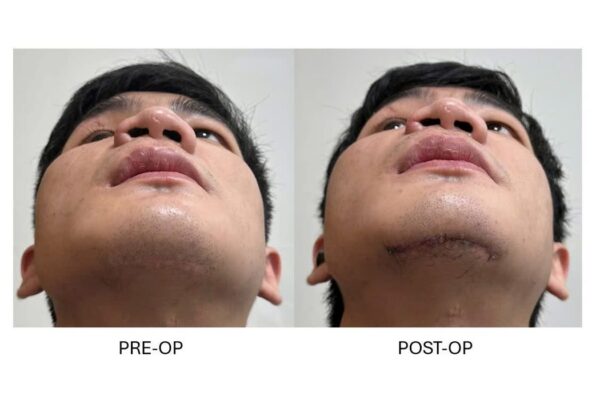
Sau khi tái tạo vùng lồi cầu, xương hàm dưới được làm dài bằng kỹ thuật kéo giãn sinh xương.
Khác với việc cắt xương rồi đưa ngay hai phân đoạn đến vị trí cuối cùng, kỹ thuật này tạo ra xương mới một cách từ từ giữa hai bề mặt xương đã được cắt.
Quá trình được thực hiện qua bốn giai đoạn.
1. Phẫu thuật:Xương hàm dưới được cắt tại vùng góc hàm hai bên và hệ thống kéo giãn Zurich II được đặt vào vị trí đã xác định.
2. Giai đoạn chờ:Sau phẫu thuật, bệnh nhân được chờ 7 ngày trước khi bắt đầu kéo giãn. Trong khoảng thời gian này, quá trình lành thương ban đầu diễn ra giữa hai bề mặt xương.
3. Giai đoạn kéo giãn:Hai phân đoạn xương được tách dần với tốc độ khoảng 1 mm mỗi ngày.
Sự kéo giãn có kiểm soát kích thích cơ thể hình thành xương mới trong khoảng trống giữa hai đầu xương. Đồng thời, các mô mềm xung quanh cũng thích nghi dần với chiều dài mới của xương hàm dưới.
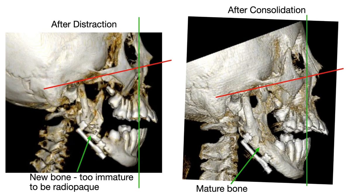
4. Giai đoạn trưởng thành xương:Sau khi đạt được chiều dài mong muốn, thiết bị được giữ nguyên để phần xương mới hình thành tiếp tục khoáng hóa và trưởng thành. Giai đoạn này thường kéo dài nhiều tháng và được theo dõi bằng thăm khám cùng hình ảnh học.
Vì sao lựa chọn kéo giãn sinh xương?
Ở một bệnh nhân 11 tuổi có xương hàm dưới rất nhỏ và cần một mức độ kéo dài đáng kể, kéo giãn sinh xương có một ưu điểm quan trọng: xương và các mô mềm liên quan được kéo dài dần dần thay vì phải thực hiện toàn bộ chuyển động trong một lần phẫu thuật.
Cùng với sự hình thành xương mới, cơ, niêm mạc, da và các mô mềm xung quanh có thời gian thích nghi với thay đổi của khung xương.
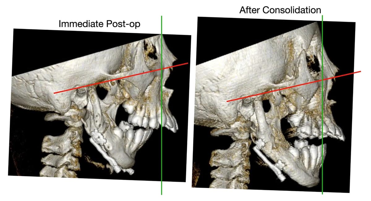
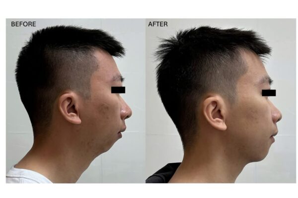
Trong trường hợp này, mục tiêu của việc đưa hàm dưới ra trước không chỉ là cải thiện hình thể khuôn mặt. Việc tăng chiều dài và đưa hàm dưới ra trước còn nhằm cải thiện tương quan của lưỡi và các cấu trúc tầng mặt dưới, qua đó góp phần giải quyết tình trạng hẹp đường thở ở bệnh nhân có ngưng thở khi ngủ tắc nghẽn.
Kết quả sau bốn tháng trưởng thành xương
Tại thời điểm đánh giá sau bốn tháng của giai đoạn trưởng thành xương, hình ảnh cắt lớp vi tính cho thấy xương mới tại vùng kéo giãn đã cốt hóa tốt.
Khả năng ăn nhai của bệnh nhân được cải thiện, đồng thời tình trạng hô hấp cũng có chuyển biến rõ rệt trên lâm sàng.
Tuy nhiên, đây chưa phải là điểm kết thúc của quá trình điều trị.
Bệnh nhân tiếp tục được điều trị chỉnh nha để giải quyết tình trạng chen chúc răng. Hàm trên cũng được nong rộng nhằm điều chỉnh bất tương xứng chiều ngang giữa hai cung hàm và có thể đồng thời cải thiện kích thước khoang mũi ở bệnh nhân phù hợp.
Điều trị sớm nhưng không phải điều trị cuối cùng
Một điểm quan trọng trong những trường hợp như thế này là cần phân biệt giữa can thiệp sớm để phục hồi chức năng và phẫu thuật chỉnh hình xương hàm cuối cùng sau khi tăng trưởng hoàn tất.
Ở tuổi 11, sự phát triển của hệ thống sọ mặt vẫn còn tiếp tục. Đặc biệt ở một bệnh nhân có bất thường bẩm sinh phức tạp và tái tạo lồi cầu bằng mảnh ghép sụn – sườn, hướng tăng trưởng trong những năm tiếp theo không thể được dự đoán hoàn toàn chính xác.
Do đó, mục tiêu hiện tại không phải là tạo ra một kết quả “hoàn chỉnh” ngay từ nhỏ.
Mục tiêu là tái lập cấu trúc cần thiết, cải thiện đường thở và chức năng, tạo thêm không gian cho hệ răng và đưa bệnh nhân đến một nền tảng thuận lợi hơn cho quá trình phát triển tiếp theo.
Khi bệnh nhân trưởng thành, sự phát triển của hai hàm và khuôn mặt sẽ được đánh giá lại. Phẫu thuật chỉnh hình xương hàm có thể được thực hiện ở giai đoạn sau để hoàn thiện tương quan hai hàm, khớp cắn và sự cân đối của khuôn mặt.
Trường hợp này cho thấy rằng tuổi còn nhỏ không phải lúc nào cũng đồng nghĩa với việc phải trì hoãn mọi can thiệp vào xương hàm. Khi biến dạng đã ảnh hưởng đáng kể đến hô hấp và chức năng, điều quan trọng là xác định đúng mục tiêu của từng giai đoạn điều trị và xây dựng một chiến lược dài hạn cho bệnh nhân.
The management of severe jaw deformities in growing children presents a fundamental question: should we intervene early, or should treatment be postponed until skeletal growth is complete?
For many dentofacial deformities, definitive orthognathic surgery is generally delayed until growth is nearly complete. However, when a skeletal deficiency is severe enough to compromise essential functions such as breathing and mastication, simply waiting for adulthood may not always be the most appropriate strategy.
This case involved an 11-year-old patient with severe mandibular hypoplasia, craniofacial asymmetry, and congenital absence of the right mandibular condyle.
Mandibular hypoplasia is more than an aesthetic problem
The patient presented with a Tessier No. 7 transverse facial cleft associated with right-sided hemifacial hypoplasia. The right mandible was severely underdeveloped, with congenital absence of the mandibular condyle.
The mandible as a whole was markedly small and retruded. This produced not only a significant facial deformity but also several functional consequences.
The underdeveloped dental arch contributed to severe dental crowding. More importantly, mandibular retrusion contributed to restriction of the retrolingual airway, and the patient was diagnosed with obstructive sleep apnea.
Treatment had previously been deferred because the patient was still growing.
In this situation, however, the objective of early intervention was not to create the patient's definitive adult facial result at the age of 11. The immediate priorities were to improve mandibular structure, breathing, and oral function while establishing a better foundation for subsequent growth and future treatment.
A staged reconstructive strategy
Following multidisciplinary assessment with Dr. Sang Nguyen Minh regarding the patient's sleep-disordered breathing, and with guidance from Professor Imad Abu El-Naaj, a staged treatment strategy was developed.
Stage 1: Condylar reconstruction
Because of the congenital absence of the right mandibular condyle, the first step was reconstruction of the condylar region using a costochondral graft.
In a growing child, a costochondral graft can be used to reconstruct the temporomandibular region and restore ramus height. Its subsequent growth behavior, however, can vary considerably between patients, making long-term surveillance throughout craniofacial development essential.
As part of the same treatment strategy, teeth 37 and 47 together with the developing 38 and 48 tooth germs were removed to facilitate the subsequent surgical and skeletal movements.
Stage 2: Mandibular distraction osteogenesis
Following reconstruction of the condylar region, the mandible was lengthened using distraction osteogenesis.
Unlike conventional osteotomy in which skeletal segments are immediately repositioned to their final location, distraction osteogenesis gradually generates new bone between surgically created bone surfaces.
The process consisted of four phases.
1. Osteotomy and distractor placement:Bilateral mandibular osteotomies were performed in the angle regions, and Zurich II distraction devices were placed.
2. Latency:A seven-day latency period was observed after surgery before activation of the distractors. During this period, the initial healing process developed between the osteotomized bone surfaces.
3. Distraction:The mandibular segments were gradually separated at approximately 1 mm per day.
Controlled mechanical distraction stimulates new bone formation within the developing gap while allowing the surrounding soft tissues to progressively adapt to the increasing mandibular length.
4. Consolidation:Once the intended advancement had been achieved, the devices were maintained in position while the newly generated bone underwent progressive mineralization and maturation. This phase extends over several months and is monitored clinically and radiographically.
Why distraction osteogenesis?
For an 11-year-old patient with a severely deficient mandible requiring substantial lengthening, distraction osteogenesis offers an important advantage: the skeletal framework and surrounding soft tissues can be lengthened progressively rather than requiring the entire correction to be achieved during a single acute movement.
As new bone forms within the distraction gap, the muscles, mucosa, skin, and other surrounding soft tissues are given time to adapt to the changing mandibular dimensions.
In this patient, mandibular advancement was intended to achieve more than an improvement in facial appearance. Increasing mandibular length and projection was also intended to improve the relationship of the tongue and lower facial structures to the upper airway, thereby contributing to management of the patient's obstructive sleep apnea.
Four months into consolidation
At four months into the consolidation phase, CT imaging demonstrated good ossification of the regenerate within the distraction sites.
The patient's masticatory function had improved, together with a clear clinical improvement in breathing.
This, however, does not represent the end of treatment.
Orthodontic treatment is continuing to address the severe dental crowding. Maxillary expansion is also being performed to correct the transverse discrepancy between the dental arches and, in appropriately selected patients, may additionally increase dimensions of the nasal cavity.
Early intervention does not mean definitive treatment
An important distinction in cases such as this is the difference between early intervention to restore function and definitive orthognathic correction after completion of growth.
At 11 years of age, substantial craniofacial growth remains. In a patient with a complex congenital deformity and a reconstructed condyle using a costochondral graft, the future growth pattern cannot be predicted with complete accuracy.
The current objective is therefore not to create a definitive adult result during childhood.
Rather, the aim is to reconstruct essential anatomy, improve the airway and function, create additional space for the developing dentition, and establish a more favorable foundation for subsequent craniofacial growth.
As the patient approaches skeletal maturity, maxillomandibular growth and facial development will be reassessed. Definitive orthognathic surgery may then be considered to further optimize jaw relationships, occlusion, and facial symmetry.
This case demonstrates that young age does not necessarily mean that all skeletal intervention should be postponed. When a severe craniofacial deformity is already compromising breathing and function, the key is to define the objective of each treatment stage and develop a long-term strategy that evolves with the patient.