Le fort iii kết hợp le fort i và bsso trong điều trị thiểu sản toàn bộ tầng mặt giữaCombined Le Fort iii, Le Fort i, and BSSO for global midface hypoplasia
02/09/2026Kết quảResults
Sai khớp cắn loại III do xương thường liên quan đến thiểu sản hàm trên, quá phát hàm dưới hoặc sự kết hợp của cả hai. Trong phần lớn trường hợp, phẫu thuật chỉnh hình xương hàm có thể được thực hiện bằng Le Fort I để đưa hàm trên ra trước, kết hợp BSSO để điều chỉnh vị trí hàm dưới và tạo hình cằm khi cần thiết.
Tuy nhiên, Le Fort I chỉ di chuyển phần hàm trên nằm bên dưới ổ mắt.
Khi biến dạng không chỉ giới hạn ở hàm trên mà liên quan đến toàn bộ tầng mặt giữa — bao gồm vùng mũi, bờ dưới ổ mắt và gò má — việc chỉ đưa phần hàm trên mang răng ra trước có thể cải thiện khớp cắn nhưng không giải quyết đầy đủ hình thể khuôn mặt.
Trường hợp này cần một chiến lược khác.
Khi Le Fort I không đủ
Bệnh nhân có sai khớp cắn loại III do xương đi kèm tình trạng thiểu sản rõ rệt của toàn bộ tầng mặt giữa.
Sự thiếu phát triển không chỉ nằm ở vùng hàm trên mà còn liên quan đến phức hợp mũi, vùng dưới ổ mắt và gò má.
Nếu chỉ thực hiện Le Fort I, phần mang răng của hàm trên có thể được đưa ra trước để cải thiện tương quan khớp cắn, nhưng các cấu trúc phía trên vẫn giữ nguyên vị trí. Điều này có thể tạo ra một tương quan xương và mô mềm chưa hài hòa dù khớp cắn đã được điều chỉnh.
Vì vậy, mục tiêu điều trị cần tách thành hai thành phần:
Le Fort III để đưa toàn bộ tầng mặt giữa ra trước.
Le Fort I để tiếp tục điều chỉnh độc lập phần hàm trên mang răng và hoàn thiện tương quan khớp cắn.
BSSO được kết hợp để đưa hàm dưới đến vị trí phù hợp với vị trí mới của hàm trên.
Vì sao Le Fort III khác với Le Fort I?
Đường cắt Le Fort III nằm cao hơn nhiều so với Le Fort I.
Phẫu thuật giải phóng phức hợp tầng mặt giữa qua vùng gốc mũi, thành và sàn ổ mắt, vùng gò má và cung tiếp, cho phép toàn bộ khối xương trung tâm của tầng mặt giữa được di chuyển như một đơn vị.
Nhờ đó, sự thay đổi không chỉ xảy ra ở vị trí răng và hàm trên mà còn có thể cải thiện độ nhô của gò má, vùng dưới ổ mắt và khung xương mũi.
Đây chính là lý do Le Fort III có vai trò trong những trường hợp thiểu sản tầng mặt giữa lan rộng mà Le Fort I đơn thuần không thể giải quyết đầy đủ.
Kết hợp Le Fort III và Le Fort I để kiểm soát riêng khuôn mặt và khớp cắn
Một lợi thế quan trọng của việc kết hợp hai mức cắt là khả năng điều chỉnh toàn bộ tầng mặt giữa và phần hàm trên mang răng tương đối độc lập với nhau.
Trước hết, Le Fort III cho phép đưa phức hợp tầng mặt giữa ra trước để tái lập hình thể vùng mũi – ổ mắt – gò má.
Sau đó, Le Fort I cho phép tinh chỉnh vị trí phần hàm trên mang răng nhằm đạt được tương quan thích hợp với cung răng hàm dưới.
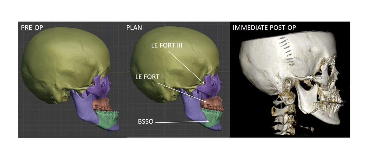
BSSO tiếp tục được sử dụng để điều chỉnh hàm dưới, tạo nên một kế hoạch ba tầng:
Le Fort III — điều chỉnh khung xương tầng mặt giữa.
Le Fort I — điều chỉnh hàm trên và khớp cắn.
BSSO — điều chỉnh vị trí hàm dưới.
Nhờ đó, phẫu thuật không phải lựa chọn giữa “khuôn mặt” và “khớp cắn”, mà có thể kiểm soát cả hai trong cùng một kế hoạch.
Lập kế hoạch ba chiều ở vùng giải phẫu gần nền sọ
Le Fort III là một phẫu thuật phức tạp vì đường cắt nằm gần nhiều cấu trúc quan trọng của ổ mắt, tầng mặt giữa và nền sọ.
Do đó, việc xác định chính xác vị trí và hướng của các đường cắt có ý nghĩa đặc biệt quan trọng.
Trong trường hợp này, toàn bộ phẫu thuật được lập kế hoạch trước trên mô hình ba chiều.
Ở vùng gốc mũi, đường cắt được thiết kế với vị trí và hướng phù hợp nhằm duy trì khoảng cách an toàn với nền sọ. Các đường cắt còn lại cũng được xây dựng dựa trên hình thái giải phẫu riêng của bệnh nhân.
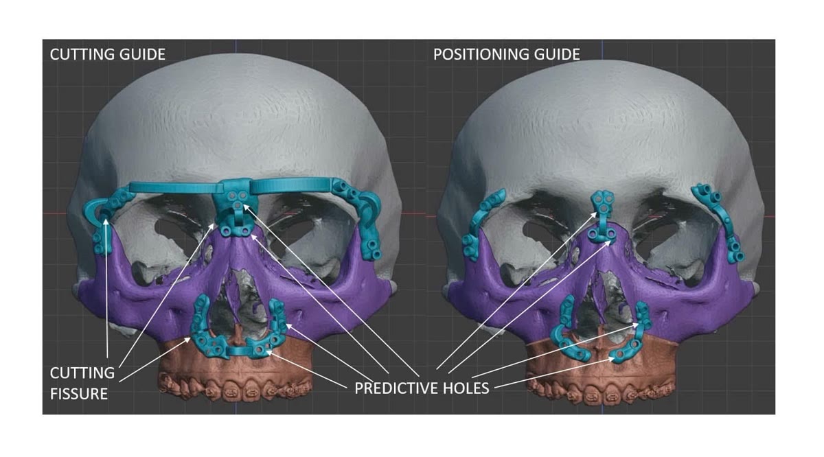
Từ kế hoạch này, máng hướng dẫn cắt xương cá nhân hóa được chế tạo để chuyển chính xác vị trí các đường cắt vào phẫu trường.
Sau khi phức hợp tầng mặt giữa được giải phóng và đưa đến vị trí mới, dụng cụ định vị cá nhân hóa được sử dụng để hỗ trợ xác định vị trí ba chiều của khối xương theo kế hoạch trước mổ.
Ở những phẫu thuật có nhiều phân đoạn và nhiều mức cắt như trường hợp này, việc kiểm soát vị trí không chỉ theo chiều trước – sau mà còn theo chiều đứng, chiều ngang và xoay của từng khối xương là yếu tố quyết định kết quả cuối cùng.
Đường tiếp cận coronal và bảo tồn tóc
Phần trên của phẫu thuật được tiếp cận thông qua đường rạch coronal trong vùng da đầu.
Đường này cung cấp phẫu trường rộng đến vùng trán, gốc mũi, ổ mắt ngoài và cung tiếp, đồng thời cho phép giấu sẹo trong vùng tóc.
Trong trường hợp này, đường rạch da đầu được thực hiện mà không cần cạo toàn bộ tóc.
Việc cầm máu bờ da đầu được thực hiện bằng phương pháp kẹp cơ học nhằm hạn chế sử dụng dao điện trực tiếp tại bờ da, qua đó giảm tổn thương nhiệt đối với các nang tóc xung quanh đường rạch.
Sau phẫu thuật, tóc tiếp tục mọc qua vùng sẹo, giúp đường mổ ngày càng khó nhận thấy khi tóc dài trở lại.
Kết quả sớm sau phẫu thuật
Toàn bộ phẫu thuật Le Fort III kết hợp Le Fort I và BSSO được hoàn thành trong khoảng bốn giờ, với lượng máu mất ước tính hơn 300 ml.
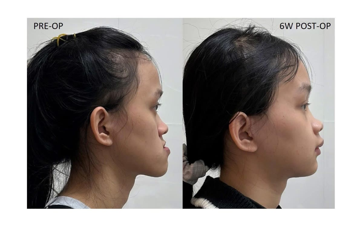
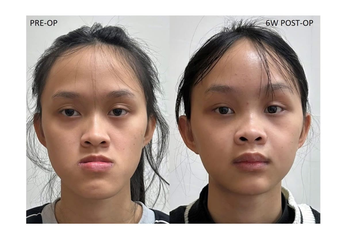
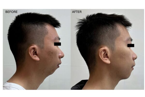
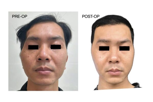
Ở thời điểm một tháng sau mổ, bệnh nhân vẫn còn phù nề sau phẫu thuật, tuy nhiên sự cải thiện về độ nhô của tầng mặt giữa đã có thể nhận thấy rõ.
Vùng mũi, dưới ổ mắt và gò má được đưa ra trước đồng thời với việc thiết lập lại tương quan hai hàm.
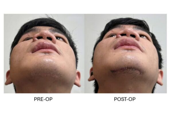
Đường rạch coronal lành tốt và tóc vẫn mọc tại vùng sẹo.
Khi vị trí khớp cắn và vị trí tầng mặt giữa cần được điều chỉnh ở hai mức khác nhau
Trường hợp này minh họa một nguyên tắc quan trọng trong phẫu thuật sọ mặt:
Mức độ của đường cắt phải phù hợp với mức độ của biến dạng.
Nếu biến dạng chỉ nằm ở hàm trên, Le Fort I có thể là lựa chọn thích hợp.
Nhưng khi toàn bộ phức hợp mũi – ổ mắt – gò má cùng bị thiểu sản, việc chỉ di chuyển phần mang răng của hàm trên sẽ không thể giải quyết đầy đủ khuôn mặt.
Trong những trường hợp được lựa chọn phù hợp, việc kết hợp Le Fort III và Le Fort I cho phép đưa toàn bộ tầng mặt giữa đến vị trí thẩm mỹ phù hợp, đồng thời vẫn kiểm soát riêng phần hàm trên mang răng để đạt được khớp cắn chính xác.
Đây cũng là giá trị của việc lập kế hoạch phẫu thuật dựa trên cấu trúc ba chiều của toàn bộ khuôn mặt, thay vì chỉ dựa vào tương quan của hai cung răng.
Skeletal Class III malocclusion commonly results from maxillary deficiency, mandibular excess, or a combination of both. In many patients, correction can be achieved using Le Fort I osteotomy for maxillary advancement, combined with bilateral sagittal split osteotomy (BSSO) to reposition the mandible and genioplasty when indicated.
However, Le Fort I moves only the tooth-bearing maxilla below the orbital level.
When the deformity involves not only the maxilla but the entire midface—including the nasal framework, infraorbital rims, and zygomatic region—advancing the Le Fort I segment alone may improve the occlusion while leaving a substantial part of the facial deficiency uncorrected.
This patient required a different strategy.
When Le Fort I is not enough
The patient presented with a skeletal Class III relationship associated with marked global midface hypoplasia.
The deficiency extended beyond the tooth-bearing maxilla to involve the nasal, infraorbital, and zygomatic components of the midface.
A conventional Le Fort I advancement could have improved the maxillomandibular relationship and occlusion, but the more superior midfacial skeleton would have remained retruded.
The treatment objectives therefore needed to be separated.
A Le Fort III osteotomy was used to advance the midfacial skeletal framework as a whole.
A Le Fort I osteotomy was then used to independently refine the position of the tooth-bearing maxilla and establish the desired occlusion.
BSSO was incorporated to position the mandible relative to the newly reconstructed maxilla.
How Le Fort III differs from Le Fort I
The Le Fort III osteotomy is performed at a substantially higher level than a Le Fort I osteotomy.
It mobilizes the midfacial skeleton through the nasofrontal region, orbital boundaries, zygomatic region, and zygomatic arches, allowing the central midface to be advanced as a single skeletal unit.
As a result, the procedure can modify not only the position of the maxillary dentition but also projection of the malar region, infraorbital skeleton, and nasal framework.
This is why Le Fort III becomes relevant in selected patients with widespread midface deficiency that cannot be adequately treated with Le Fort I advancement alone.
Combining Le Fort III and Le Fort I for independent control of the face and occlusion
One of the principal advantages of combining these two osteotomy levels is the ability to control the global midface and the tooth-bearing maxilla relatively independently.
The Le Fort III movement first establishes the desired position of the overall midfacial framework, restoring projection of the nasal–orbital–zygomatic complex.
The Le Fort I segment can then be independently adjusted to refine maxillary position and achieve the required relationship with the mandibular dentition.
BSSO is subsequently used to reposition the mandible.
The operation therefore becomes a three-level strategy:
Le Fort III — correction of the global midfacial skeleton.
Le Fort I — refinement of the maxilla and occlusion.
BSSO — correction of mandibular position.
This allows facial projection and dental occlusion to be treated as related but not identical surgical objectives.
Three-dimensional planning close to the skull base
Le Fort III osteotomy is technically demanding because the osteotomy lines are located close to important orbital, midfacial, and skull-base structures.
Precise determination of both the location and orientation of these osteotomies is therefore essential.
For this patient, the entire procedure was planned preoperatively in a three-dimensional virtual environment.
At the nasofrontal region, the osteotomy was designed at an appropriate level and trajectory to maintain a safe relationship with the anterior skull base. The remaining osteotomy lines were similarly adapted to the patient's individual skeletal anatomy.
Based on this plan, patient-specific cutting guides were manufactured to transfer the designed osteotomies to the surgical field.
Following mobilization and advancement of the midfacial segment, patient-specific positioning devices were used to assist in reproducing its planned three-dimensional position.
In an operation involving multiple osteotomy levels and independently mobile skeletal segments, control is required not only in the anteroposterior direction but also in the vertical, transverse, and rotational dimensions.
Coronal exposure and preservation of the hair-bearing scalp
The superior component of the procedure was approached through a coronal scalp incision.
This provides wide exposure of the frontal region, nasofrontal area, lateral orbit, and zygomatic arches while allowing the final scar to remain concealed within the hair-bearing scalp.
In this patient, complete shaving of the scalp was not required.
Mechanical edge control was used during the scalp incision to assist hemostasis while limiting direct thermal injury from electrocautery near the incision margins and adjacent hair follicles.
During postoperative healing, hair continued to grow through the scar region, making the incision increasingly inconspicuous as the hair lengthened.
Early postoperative result
The combined Le Fort III, Le Fort I, and BSSO procedure was completed in approximately four hours, with estimated blood loss slightly above 300 mL.
At one month after surgery, postoperative edema was still present, but improvement in midfacial projection was already evident.
The nasal, infraorbital, and zygomatic regions had been advanced together while the maxillomandibular relationship was simultaneously reconstructed.
The coronal incision healed well, with preserved hair growth across the scar region.
Matching the osteotomy level to the level of the deformity
This case illustrates a fundamental principle in craniofacial surgery:
The level of the osteotomy should correspond to the level of the deformity.
When the deficiency is limited primarily to the maxilla, Le Fort I advancement may be sufficient.
When the entire nasal–orbital–zygomatic complex is retruded, however, repositioning only the tooth-bearing maxilla cannot fully correct the facial deficiency.
In appropriately selected patients, combining Le Fort III with Le Fort I allows the surgeon to advance the global midface to the desired facial position while independently controlling the tooth-bearing maxilla to achieve an accurate occlusal relationship.
The case therefore demonstrates the value of planning orthognathic and craniofacial surgery around the three-dimensional architecture of the entire face rather than the dental arches alone.