Lõm mắt sau chấn thương: tái tạo ổ mắt trong trường hợp điều trị muộnPost-traumatic enophthalmos: secondary orbital reconstruCTion
02/09/2026Kết quảResults
Lõm mắt (enophthalmos) là một trong những di chứng có thể gặp sau chấn thương ổ mắt, đặc biệt khi tổn thương sàn hoặc thành ổ mắt không được phát hiện và tái tạo đầy đủ trong giai đoạn ban đầu.
Ở bệnh nhân chấn thương, lõm mắt không đơn thuần là vấn đề của nhãn cầu mà thường liên quan trực tiếp đến sự thay đổi cấu trúc ba chiều của toàn bộ ổ mắt.
Tại sao chấn thương ổ mắt có thể gây lõm mắt?
Ổ mắt là một cấu trúc xương ba chiều có hình thái phức tạp. Khi sàn ổ mắt hoặc thành trong ổ mắt bị vỡ, giới hạn xương của ổ mắt bị thay đổi, làm tăng thể tích khoang ổ mắt.
Đồng thời, mô mềm trong ổ mắt có thể thoát vị hoặc di lệch qua vùng xương bị khuyết vào xoang hàm trên hoặc xoang sàng.
Sự kết hợp giữa tăng thể tích khoang ổ mắt và thay đổi vị trí của mô mềm ổ mắt làm nhãn cầu dịch chuyển ra phía sau, tạo nên tình trạng lõm mắt.
Trong một số trường hợp, teo mô mỡ ổ mắt sau chấn thương cũng có thể góp phần vào biến dạng, nhưng đây không phải là cơ chế duy nhất.
Chấn thương ổ mắt có thể xuất hiện đơn độc hoặc phối hợp với các dạng chấn thương phức tạp hơn như gãy phức hợp gò má – hàm trên (ZMC), gãy xương hàm trên hoặc gãy phức hợp mũi – ổ mắt – sàng (NOE).
Khi mức độ lõm mắt đủ lớn, đặc biệt khoảng trên 2 mm, sự khác biệt giữa hai mắt có thể trở nên rõ ràng và ảnh hưởng đáng kể đến thẩm mỹ khuôn mặt.
Một trường hợp lõm mắt sau chấn thương điều trị muộn
Bệnh nhân nữ, 25 tuổi, trước đó đã được điều trị chấn thương vùng mặt tại một cơ sở y tế khác.
Sau giai đoạn điều trị ban đầu, bệnh nhân đến Bệnh viện Răng Hàm Mặt Trung ương Hà Nội để tháo phương tiện kết hợp xương và đồng thời mong muốn điều trị tình trạng lõm mắt còn tồn tại.
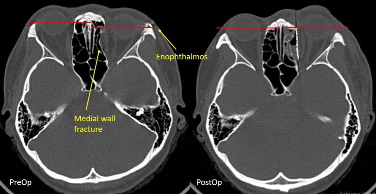
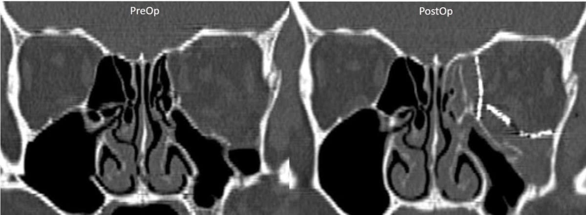
Hình ảnh CT cho thấy đây là một tổn thương phức tạp, với khuyết xương liên quan đồng thời đến sàn và thành trong ổ mắt.
Một khó khăn khác là tổn thương đã tồn tại trong thời gian dài.
Ở giai đoạn muộn, các mô mềm ổ mắt có thể đã hình thành xơ dính và thích nghi với vị trí bất thường; các mốc giải phẫu cũng trở nên khó xác định hơn so với chấn thương mới. Vì vậy, việc tái lập chính xác hình thái ba chiều của ổ mắt trở nên khó khăn hơn đáng kể.
Vì sao thời điểm điều trị quan trọng?
Không phải tất cả các trường hợp vỡ sàn hoặc thành ổ mắt đều cần phẫu thuật và thời điểm can thiệp cần được quyết định dựa trên nhiều yếu tố như thị lực, vận nhãn, song thị, mức độ kẹt mô, kích thước khuyết xương và sự thay đổi vị trí nhãn cầu.
Tuy nhiên, ở những trường hợp có chỉ định tái tạo ổ mắt, việc phát hiện và xử trí phù hợp trong giai đoạn sớm thường thuận lợi hơn so với tái tạo một biến dạng đã tồn tại lâu ngày.
Khi bệnh nhân đến ở giai đoạn muộn với tình trạng lõm mắt đã hình thành, phẫu thuật trở thành một secondary orbital reconstruction – tái tạo ổ mắt thứ phát, với mức độ phức tạp cao hơn đáng kể.
Tái tạo lại cấu trúc ba chiều của ổ mắt
Mục tiêu của phẫu thuật không đơn thuần là “đẩy mắt ra phía trước”.
Điều quan trọng là phải xác định được cấu trúc xương đã mất hoặc biến dạng, giải phóng các mô mềm bị di lệch và tái lập lại hình thái cũng như thể tích của ổ mắt gần với giải phẫu ban đầu nhất có thể.
Đây cũng là lý do việc đánh giá CT và hiểu rõ giải phẫu ba chiều của ổ mắt đóng vai trò đặc biệt quan trọng trong những trường hợp tái tạo muộn.
Trong quá trình đào tạo và làm việc tại Nhật Bản, mình có cơ hội tiếp xúc và điều trị nhiều trường hợp chấn thương ổ mắt. Những kinh nghiệm này, kết hợp với các kiến thức cập nhật từ các hội nghị quốc tế về chấn thương và tái tạo vùng hàm mặt, đã hỗ trợ mình rất nhiều khi xử lý trường hợp phức tạp này.
Sau phẫu thuật tái tạo, tình trạng lõm mắt của bệnh nhân đã cải thiện rõ rệt. Tuy nhiên, với những tổn thương ổ mắt phức tạp đã tồn tại lâu ngày, mục tiêu điều trị cần thực tế: cải thiện tối đa hình thái, vị trí nhãn cầu và sự cân đối giữa hai bên, thay vì kỳ vọng có thể khôi phục hoàn toàn cấu trúc như trước chấn thương.
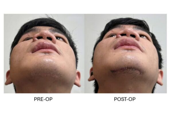
Hình ảnh trong bài minh họa CT của bệnh nhân trước và sau phẫu thuật tái tạo ổ mắt.
Enophthalmos, or posterior displacement of the globe, is an important potential sequela of orbital trauma, particularly when fractures involving the orbital floor or walls are not adequately identified and reconstructed during the initial treatment.
Post-traumatic enophthalmos is not simply a problem of the globe itself. It is frequently the result of alteration of the complex three-dimensional anatomy of the orbit.
Why can orbital trauma cause enophthalmos?
The orbit is a complex three-dimensional bony structure. When the orbital floor or medial wall is fractured, disruption of the bony boundaries can increase the volume of the orbital cavity.
At the same time, orbital soft tissues may herniate or become displaced through the defect into the maxillary or ethmoid sinuses.
The combination of increased bony orbital volume and displacement of orbital soft tissues can result in posterior displacement of the globe and clinically apparent enophthalmos.
Post-traumatic atrophy of orbital fat may also contribute to the deformity in some patients, but it represents only one component of a multifactorial process.
Orbital fractures may occur in isolation or as part of more extensive facial injuries, including zygomaticomaxillary complex (ZMC) fractures, maxillary fractures, and naso-orbito-ethmoidal (NOE) fractures.
When enophthalmos becomes sufficiently pronounced, particularly at approximately 2 mm or greater, the asymmetry between the two eyes may become clinically noticeable and have a significant aesthetic impact.
A challenging case of delayed post-traumatic enophthalmos
A 25-year-old female patient had previously undergone treatment for facial trauma at another institution.
She subsequently presented to the National Hospital of Odonto-Stomatology, Hanoi, for removal of her osteosynthesis hardware and requested correction of persistent post-traumatic enophthalmos.
CT examination demonstrated a complex orbital defect involving both the orbital floor and medial wall.
An additional challenge was the delayed presentation.
In secondary cases, displaced orbital tissues may have developed fibrosis and adhesions, while normal anatomical landmarks can be more difficult to identify than in an acute fracture. Consequently, accurate restoration of the three-dimensional orbital anatomy becomes considerably more challenging.
Why does timing matter?
Not every orbital floor or wall fracture requires surgical reconstruction, and the timing of intervention must be determined according to multiple clinical factors, including visual function, ocular motility, diplopia, tissue entrapment, defect size, and globe position.
However, when orbital reconstruction is indicated, appropriate assessment and management during the earlier stages of injury are generally more favorable than correction of an established late deformity.
Once post-traumatic enophthalmos has developed, treatment becomes a secondary orbital reconstruction, which is technically considerably more demanding.
Restoring the three-dimensional anatomy of the orbit
The objective of orbital reconstruction is not simply to “push the eye forward.”
The fundamental goal is to identify the disrupted or missing bony anatomy, release displaced soft tissues, and restore the shape and volume of the orbit as closely as possible to its pre-injury anatomy.
This is why careful CT assessment and a detailed understanding of three-dimensional orbital anatomy are particularly important in delayed orbital reconstruction.
During my surgical training and clinical work in Japan, I had the opportunity to gain substantial exposure to the assessment and management of orbital trauma. This experience, together with knowledge acquired through international meetings focusing on maxillofacial trauma and reconstruction, was particularly valuable in the management of this challenging case.
Following secondary orbital reconstruction, the patient's enophthalmos showed significant improvement.
Nevertheless, in complex orbital injuries treated at a delayed stage, expectations must remain realistic. The objective is to achieve the greatest possible restoration of orbital anatomy, globe position, and facial symmetry rather than to assume that the original pre-traumatic anatomy can always be completely reproduced.
The images presented demonstrate the patient's CT findings before and after secondary orbital reconstruction.