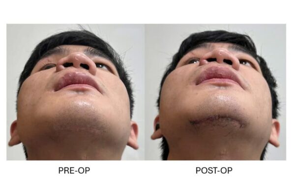
Tái tạo chấn thương phức hợp mũi – sàng – ổ mắt hai bênReconstruCTion of complex bilateral nASO-orbito-ethmoid and orbital trauma
02/09/2026Kết quảResults
Chấn thương phức hợp mũi – sàng – ổ mắt là một trong những dạng chấn thương phức tạp của tầng mặt giữa. Tổn thương không chỉ gây biến dạng vùng mũi mà còn có thể làm thay đổi hình dạng và thể tích ổ mắt, ảnh hưởng đến vị trí nhãn cầu, hệ thống lệ và trong những trường hợp nặng có thể phối hợp với tổn thương nền sọ.
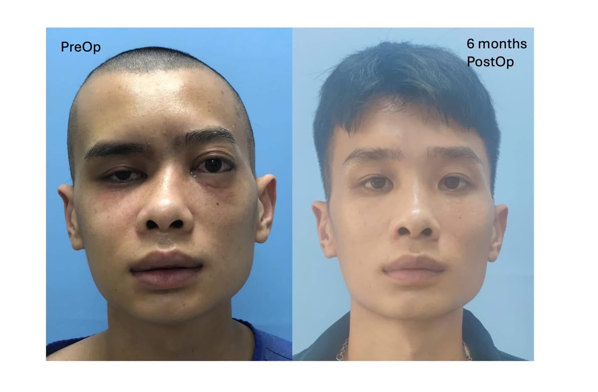
Trường hợp này là một bệnh nhân nam, 24 tuổi, bị đa chấn thương vùng sọ mặt sau tai nạn giao thông, bao gồm vỡ thành trước xoang trán, gãy phức hợp mũi – sàng – ổ mắt hai bên và gãy phức hợp gò má bên phải.
Khi một khối xương trung tâm làm biến dạng cả hai ổ mắt
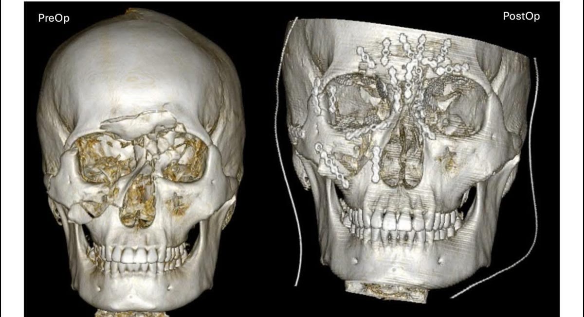
Sau chấn thương, khối xương mũi – sàng ở trung tâm khuôn mặt bị ép lùi ra sau và lệch sang trái.
Sự dịch chuyển này tạo ra những biến đổi khác nhau ở hai bên ổ mắt.
Ở bên phải, ổ mắt bị mở rộng, làm tăng thể tích ổ mắt và góp phần khiến nhãn cầu tụt về phía sau, gây lõm mắt.
Ngược lại, phía bên trái bị thu hẹp không gian ổ mắt, khiến nhãn cầu bị đẩy ra trước tương đối.
Kết hợp với tổn thương xương gò má phải, toàn bộ vùng trung tâm và tầng mặt trên của bệnh nhân bị biến dạng rõ rệt.
Tổn thương còn phức tạp hơn do trần ổ mắt hai bên bị vỡ nhiều mảnh và thông với vùng nền xoang trán.
Vì vậy, mục tiêu phẫu thuật không đơn thuần là cố định từng đường gãy riêng lẻ mà phải tái lập lại cấu trúc ba chiều của toàn bộ vùng trán – mũi – sàng – ổ mắt – gò má.
Tái lập khung xương trước khi tái tạo ổ mắt
Phẫu thuật được thực hiện thông qua đường rạch coronal hai bên, cho phép bộc lộ rộng vùng trán, bờ trên ổ mắt, phức hợp mũi – sàng và cung tiếp – gò má.
Các cấu trúc xương bị di lệch, bao gồm khối mũi – sàng, thành trước xoang trán và phức hợp gò má phải, được đưa trở lại vị trí giải phẫu và cố định bằng nẹp vít.
Việc phục hồi chính xác khung xương xung quanh ổ mắt là bước đặc biệt quan trọng.
Ổ mắt là một cấu trúc ba chiều phức tạp. Nếu các bờ và khung xương xung quanh chưa được đưa về đúng vị trí mà đã tiến hành tái tạo thành ổ mắt, hình dạng và thể tích ổ mắt sau cùng có thể vẫn không chính xác.
Sau khi khung xương được tái lập, các khuyết hổng thành ổ mắt được tái tạo bằng lưới titan nhằm phục hồi hình dạng và thể tích ổ mắt gần với giải phẫu ban đầu nhất có thể.
Mục tiêu cuối cùng là tái lập đồng thời hình dạng tầng mặt giữa, sự cân đối của hai ổ mắt và vị trí của nhãn cầu.
Vì sao gãy phức hợp mũi – sàng – ổ mắt cần được đánh giá cẩn thận?
Tổn thương vùng mũi – sàng không chỉ gây bẹt hoặc biến dạng sống mũi.
Đây là khu vực giải phẫu nằm tại giao điểm giữa mũi, ổ mắt, xoang trán và nền sọ, vì vậy chấn thương năng lượng cao có thể ảnh hưởng đến nhiều cấu trúc quan trọng.
Sự thay đổi vị trí của thành trong ổ mắt có thể làm thay đổi thể tích ổ mắt và vị trí nhãn cầu. Hệ thống dẫn lưu lệ cũng có thể bị tổn thương. Khi đường gãy lan đến nền sọ, bệnh nhân có nguy cơ rò dịch não tủy và các biến chứng nội sọ.
Trong chấn thương ổ mắt nói chung, những biến chứng cấp tính như tụ máu sau nhãn cầu kèm tăng áp lực ổ mắt cũng cần được nhận biết nhanh chóng vì có thể đe dọa thị lực nếu không được xử trí kịp thời.
Do đó, việc đánh giá một bệnh nhân có chấn thương phức tạp vùng mũi – ổ mắt không nên chỉ tập trung vào hình dạng của mũi hoặc các đường gãy nhìn thấy rõ, mà cần xem xét toàn bộ cấu trúc sọ – mặt liên quan.
Vai trò của đường tiếp cận coronal
Đường coronal là một đường tiếp cận kinh điển trong phẫu thuật sọ mặt, cho phép bộc lộ rộng nhiều vùng giải phẫu thông qua một đường rạch được giấu trong vùng da đầu.
Tùy theo phạm vi bóc tách, đường tiếp cận này có thể tạo phẫu trường thuận lợi đến xương trán, bờ trên và thành ngoài ổ mắt, phức hợp mũi – sàng, cung tiếp và vùng gò má.
Trong những chấn thương phức tạp như trường hợp này, khả năng quan sát đồng thời nhiều cấu trúc giúp phẫu thuật viên tái lập toàn bộ khung xương sọ mặt theo một hệ thống thống nhất, thay vì tiếp cận từng đường gãy qua nhiều đường rạch nhỏ riêng biệt.
Đường tiếp cận vùng cung tiếp cũng cần được lựa chọn dựa trên giải phẫu của nhánh trán thần kinh mặt. Tổn thương nhánh thần kinh này có thể gây yếu hoặc liệt vận động vùng trán. Vì vậy, hiểu rõ các lớp giải phẫu và lựa chọn mặt phẳng bóc tách phù hợp là yếu tố quan trọng khi tiếp cận vùng thái dương – cung tiếp.
Tái tạo chấn thương sọ mặt là tái tạo một cấu trúc ba chiều
Trường hợp này minh họa một nguyên tắc quan trọng trong điều trị chấn thương sọ mặt phức tạp:
Không thể tái tạo chính xác ổ mắt nếu khung xương bao quanh ổ mắt chưa được phục hồi chính xác.
Một vài milimét sai lệch của vùng mũi – sàng, bờ ổ mắt hoặc xương gò má có thể làm thay đổi hình dạng và thể tích ổ mắt, từ đó ảnh hưởng đến vị trí nhãn cầu và sự cân đối của khuôn mặt.
Vì vậy, với những chấn thương phức tạp vùng mũi – sàng – ổ mắt, mục tiêu của phẫu thuật không chỉ là “nắn và cố định đường gãy”, mà là tái tạo lại cấu trúc ba chiều của toàn bộ vùng sọ mặt bị tổn thương.
Naso-orbito-ethmoid (NOE) fractures are among the more complex injuries of the central midface. Beyond nasal deformity, displacement of the central facial skeleton can alter orbital shape and volume, affect globe position, injure the lacrimal drainage system, and, in severe trauma, occur in association with anterior skull-base injuries.
This case involved a 24-year-old man who sustained extensive craniofacial trauma following a road traffic accident, including an anterior frontal sinus wall fracture, bilateral NOE fractures, and a right zygomaticomaxillary complex fracture.
When central facial displacement affects both orbits differently
Following the injury, the central nasoethmoidal complex was displaced posteriorly and toward the left.
This produced different effects on the two orbits.
On the right side, displacement of the orbital framework resulted in enlargement of the orbit, contributing to posterior displacement of the globe and post-traumatic enophthalmos.
On the left side, the orbital space became relatively narrowed, resulting in relative anterior displacement of the globe.
Together with the right zygomatic injury, these changes produced severe distortion of the central and upper facial skeleton.
The injury was further complicated by comminuted fractures of both orbital roofs communicating with the frontal sinus region.
The reconstructive objective was therefore not simply to fix individual fracture lines, but to restore the three-dimensional architecture of the entire frontal–nasoethmoidal–orbital–zygomatic complex.
Reconstructing the facial framework before reconstructing the orbit
Surgical exposure was obtained through a bilateral coronal approach, providing wide access to the frontal bone, superior orbital rims, nasoethmoidal region, and zygomatic arches.
The displaced skeletal components—including the nasoethmoidal complex, anterior frontal sinus wall, and right zygomatic complex—were repositioned toward their anatomical locations and stabilized with internal fixation.
Accurate restoration of the skeletal framework surrounding the orbit was a critical step.
The orbit is a complex three-dimensional structure. If its surrounding boundaries are not correctly repositioned before orbital wall reconstruction, the final orbital shape and volume may remain inaccurate even when the wall defect itself is adequately covered.
Once the surrounding framework had been restored, the orbital defects were reconstructed with titanium mesh to reproduce the original orbital anatomy and volume as closely as possible.
The overall objective was to simultaneously restore midfacial architecture, orbital symmetry, and globe position.
Why NOE injuries require careful assessment
NOE trauma can produce much more than flattening or displacement of the nasal bridge.
The nasoethmoidal region lies at the anatomical intersection of the nose, orbits, frontal sinus, and anterior skull base. High-energy trauma in this area can therefore involve several functionally important structures.
Displacement of the medial orbital walls can alter orbital volume and globe position. The lacrimal drainage apparatus may be injured. Extension of the fracture toward the skull base may be associated with cerebrospinal fluid leakage and intracranial complications.
In orbital trauma more broadly, acute complications such as retrobulbar hemorrhage with orbital compartment syndrome must also be recognized promptly because delayed treatment can result in permanent visual loss.
Assessment of complex central facial trauma should therefore extend beyond the obvious nasal deformity or individual fracture lines and consider the entire craniofacial injury pattern.
The role of the coronal approach
The coronal approach is a classic craniofacial surgical exposure that provides broad access through an incision concealed within the scalp.
Depending on the extent and plane of dissection, it can provide excellent exposure of the frontal bone, superior and lateral orbital regions, nasoethmoidal complex, zygomatic arches, and adjacent craniofacial skeleton.
In extensive injuries such as this one, wide exposure allows multiple displaced skeletal components to be visualized simultaneously and reconstructed as part of a single anatomical framework rather than treating each fracture independently through multiple limited incisions.
Surgical access to the zygomatic arch and temporal region also requires careful consideration of the anatomy of the frontal branch of the facial nerve. Injury to this branch may result in weakness of forehead movement. Appropriate surgical planes and detailed knowledge of the temporal anatomy are therefore essential when exposing this region.
Craniofacial trauma reconstruction is a three-dimensional problem
This case illustrates a fundamental principle of complex craniofacial trauma reconstruction:
The orbit cannot be accurately reconstructed until the skeletal framework surrounding it has been accurately restored.
Even a few millimeters of residual displacement involving the nasoethmoidal complex, orbital rims, or zygomatic skeleton can alter orbital geometry and volume, potentially affecting globe position and facial symmetry.
For this reason, the objective in complex NOE and orbital trauma extends beyond simply reducing and fixing individual fractures. The ultimate goal is to reconstruct the three-dimensional architecture of the injured craniofacial skeleton as a whole.